Professor Mark Evans explains his research into cellular networks that can be rapidly dismantled, reconfigured and rebuilt.
Our understanding of cells and the way they work has taken a significant step forward, with research that has found an internal communication system manages the signals that tell a cell what to do and can quickly reconfigure itself to tell the cell to do something different.
The researchers, headed by Professor Mark Evans in the University of Edinburgh’s Centre for Discovery Brain Sciences, have dubbed this signalling network “the cell-wide web”. They claim that their work rewrites the book on how instructions spread around a cell, and that deciphering the deeper code that controls this cell-wide web could lead to more targeted therapies for conditions such as cancer and hypertension.
Conventional wisdom
The discovery did not happen spontaneously or in isolation. In fact, says Mark Evans, its foundations were laid during the 1970s: “The hint of it emerged when the Dutch-Canadian scientist Casey van Breemen suggested the presence of what he called the ‘superficial buffer barrier’, which would restrict calcium movement across the very small space between the membrane of the cell and the membrane of the major internal calcium store, an organelle known as the sarco/endoplasmic reticulum. Effectively, his experiments predicted that calcium went straight into the sarcoplasmic reticulum and not into the general cytoplasm.”
Conventional wisdom had said that the signals instructing a cell travelled in waves through the gel-like cytoplasm. But van Breemen’s ideas questioned this, as did Evans’ work in the early 2000s. “My experiments were inconsistent with the cell being an open sea of cytoplasm,” he says. “But we couldn’t see anything. We had just a few images with poor spatial resolution. Our studies consistently suggested their function was coordinated by more complex and site-specific calcium signaling.” Evans’ work also predicted that intracellular signalling was coordinated by a wider array of junctional complexes, through a system with similar stimulus-response characteristics to the neuromuscular junction, which guides the action of transmitters released by nerve impulses.
Collaboration
Then, in 2005, Evans and van Breemen (now working in Canada) ran into each other at a symposium in Oxford. “He knew about my research and said we should collaborate, even though at that point I didn’t know who he was or what he did,” says Evans. Via Skype, they discussed ways to explore calcium signalling across the superficial buffer barrier, and deliberated over the expanding number of intracellular junctions that appeared to have a width
of less than one ten-thousandth of a millimetre. Computer models confirmed that the junctions between cell membranes would only operate effectively on the nano-scale, likely below 100nm, throwing more light on the role these might play in directing signals. But, at this point, anybody wanting to see the junctions in action would have to wait for the technology to catch up.
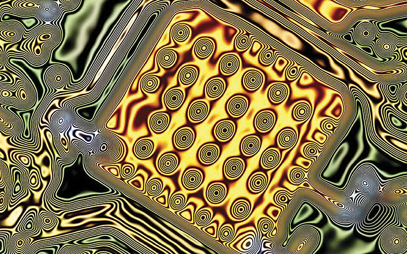
This finally happened in 2012 when a new microscopy system from Nikon appeared in the University of Edinburgh research labs. “We were now able to accurately acquire complete time series
of images of very thin whole sections through a cell,” says Evans. “We could visualise calcium signals within small tributaries of cytoplasm segregated from the rest of the cell by invaginations of the nuclear-envelope membranes, forming tubes rather than single-plane junctional complexes and offering even greater capacity to direct very local activity. In short, these invaginations project into the nucleus and carry some cytoplasm in the middle of them, so it’s a segregated space that provides a distinct area of cytoplasm that can receive signals separate from the rest of the cell.”
The team then ran algorithms to clean up the image and remove any out-of-focus light. This revealed that the tubes were not only in the nucleus but extended throughout the entire cell – in doing so, they visualised for the first time the superficial buffer barrier, just as van Breemen, now in his 80s, predicted in 1977.
Changing network
Another discovery followed. “When we cultured cells the network completely changed,” says Evans. “So a growing cell is configured in a different way, and signals in a very different way, than a cell that is differentiated and carrying out the function it was designed to do.”
In terms of understanding this, Evans draws an analogy with the internet. “Think of the nerves coming from our brains to our muscles as our internet, and when we think, ‘lift our arm up,’ the muscle does that through the neuromuscular junction. What we have discovered is a cellular intranet that functions in a similar manner. The tubules control specific areas of a cell to determine what they do. For example, in a muscle cell, you can make it relax or contract. If you relax it lowers blood pressure, and if it contracts you raise blood pressure. So, if we can understand better the arrangement of the machinery inside the cell, then we can target it to raise or lower blood pressure.”
Closer investigation of nuclear invaginations has also yielded the first visual insight into how small molecule drugs might regulate gene expression and adjust cell function that is abnormal at the genetic level. Pharmaceutical companies have been developing drugs that change gene expression, but only now can they actually see how they might work via the regulatory system of the nuclear invaginations. “We are the first to ascribe a physiological function to nuclear invaginations,” says Evans, “and to show they are the site for the regulation of gene expression on a minute-to-minute basis.”
The priority now for Evans and his team is to characterise the protein complexes that guide signals to different parts of the cell. “On top of that, we want to understand how this network can be dismantled, reconfigured and rebuilt,” he says. “This is something totally new, and I never thought for one minute it would be as a complex as this!”
Mark Evans
- 1990-94: postdoctoral research, University of Edinburgh and University of London
- 1994-96: visiting research fellow, University of London
- 1996-2001: research fellow, University of Oxford
- 2001-07: lecturer and reader, University of St Andrews
- 2007-08: professor, cell biology, University of East Anglia
- 2008 - present: senior academic fellow, University of Edinburgh
- 2009 - present: chair of Cellular Pharmacology, University of Edinburgh