Schistosomiasis, a parasitic disease affecting millions worldwide, poses a significant public health and economic burden, particularly in impoverished regions.
Currently, microscopy is the standard for diagnosing schistosomiasis, but it is time-consuming, operator-dependent, and requires specialised expertise, making it challenging for resource-limited areas.
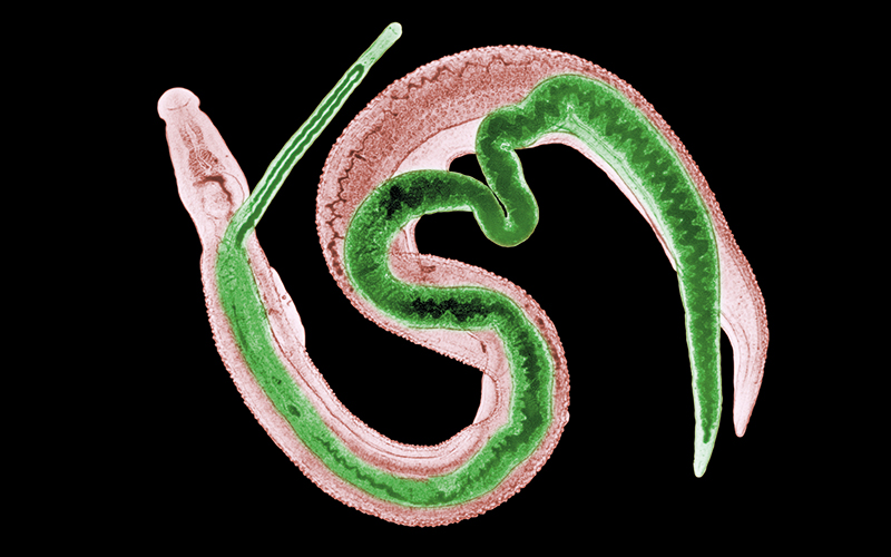
Researchers have now developed the Schistoscope – an innovative optical tool equipped with an autofocusing and automated slide-scanning system. This device captures microscopy images of urine samples, enabling efficient detection of Schistosoma haematobium (SH) eggs, a common cause of urogenital schistosomiasis.
The team created an SH dataset consisting of 12,051 images of urine samples collected in a rural area in central Nigeria and captured using the Schistoscope device.
They then manually annotated the images, marking the eggs and differentiating them from artefacts, such as crystals, glass debris, air bubbles, and fibres, which can hinder diagnosis.
In the first stage, the framework performs semantic segmentation to identify candidate SH eggs in the captured images. The second stage refines the segmentation by fitting overlapping ellipses, effectively separating boundaries of clustered eggs, leading to more accurate egg counts.
The researchers implemented it on an edge AI system and tested it on 65 clinical urine samples obtained in a field setting in Nigeria. The results showed high sensitivity, specificity and precision, with the automated egg count closely correlated to the manual count by an expert microscopist.
Image Credit | Science-Photolibrary