The Lutheran blood group system (BGS) consists of a large number of antigens, 24 being recognised by the International Society of Blood Transfusion at the time of writing, and is also of interest in terms of the various backgrounds to the (apparent) Lu(a-b-) phenotypes. The Lutheran BGS is also incorrectly named, as will be explained shortly.
There are four antithetical pairs of antigens within the BGS, but certain antigens (for example, Lu10) appear to be missing. This is the result of so-called para-Lutheran antigens, reported as possibly belonging to the Lutheran BGS but later proved to belong elsewhere.
Most of the Lutheran BGS antigens are high prevalence, with Lua, Aua and Aub being polymorphic and only Lu9 and Lu14 being low prevalence.
Lutheran antigens and antibodies tend to result in agglutination that gives a characteristic loose agglutination, with many unagglutinated red cells. An expert and experienced blood group serologist can sometimes use this characteristic agglutination to guide them towards recognising an antibody directed against an antigen within the Lutheran BGS.
Nomenclature
The first antigen described within this particular BGS, Lua, was described briefly by Callender et al in 1945, and in more detail by Callender and Race in 1946. The cognate antibody was discovered in a patient with lupus erythematosus, who also produced the first described anti-Cw of the Rh BGS and anti-Kpc of the Kell BGS.
In those days, the antibody tended to be named after the donor who had stimulated the antibody. In this case, the w of Cw stands for the donor Willis. Anti-Kpc was originally named after the donor Levay, an antibody directed against an extremely low prevalence antigen that was only recognised to be part of the Kell BGS much later in 1979. Anti-Lua was named after the donor Lutheran – except that the donor was not actually named Lutheran, but was in fact called Lutteran. However, the handwriting of the doctor who had taken the donor’s blood had been misinterpreted (plus ça change, plus c’est la même chose!), and this is why I said earlier that the Lutheran BGS had been incorrectly named.
The antithetical antigen, Lub, and its cognate antibody were described by Cutbush and Chanarin in 1956.
For a while, an alpha-numeric system was used to name antigen and antibodies (Aua and Aub are “trivial names” for Lu18 and Lu19 respectively, but were originally thought to be the only two members of an independent BGS named Auberger, and the trivial names have stuck). Now the custom is to use LU to signify that the antigen belongs to the Lutheran BGS, and the third and fourth letters denote the change in amino acid residue from the “wild-type” carrier molecule (hence LURC has a change from arginine [R] to cysteine [C] in the antigen negative type, at position 75).
Lu(a-b-) phenotypes
In 1961, Crawford et al described a sample of blood that was apparently Lu(a-b-). This proved to be unusual in two ways. Firstly, this apparently Lu(a-b-) individual was Crawford herself; secondly, the red cells were not actually Lu(a-b-), but expressed very low levels of Lutheran BGS antigens (by adsorption and elution), but were inhibited from expressing normal levels of these antigens by a dominant inhibitor gene, located on a chromosome different from the chromosome where the antigens of the Lutheran BGS are encoded (chromosome 19). In the case of an In(Lu) type of apparent Lu(a-b-), as this is called, the expression of some other antigens are also weakened, notably AnWj, P1, I, CD44 (Inb), those of the Knops BGS, Csa (disputed) and MER2, while the expression of CD75 may be elevated.
In 1963, Darnborough et al described an Lu(a-b-) individual who had what they then called anti-Luab and what is now known as anti-Lu3. This individual was genuinely Lu(a-b-).
In 1986, Norman et al described another form of apparent Lu(a-b-), this time in an Australian family that showed the cause to be X-linked (this was revealed when five other members of the family were found to be apparently Lu(a-b-), and these were all males). Once again, weak expression of the Lutheran antigens could be demonstrated by adsorption and elution and, in this case, weak expression of I, and enhanced expression of i. This type has been found in just this one family.
Only the true amorphic form of Lu(a-b-) can make anti-Lu3, but such individuals can tolerate blood from other individuals with the amorphic form of Lu(a-b-), but also from individuals with the In(Lu) type of apparent Lu(a-b-) – and, presumably, from individuals with the X-linked type of apparent Lu(a-b-).
Carrier molecules
There are two slightly different carrier molecules, both of which are described as being part of the immunoglobulin superfamily (IgSF). Each is made up of three constant regions and two variable regions. The first, the Lutheran glycoprotein is of apparent molecular weight of 85kDa, while the second, the basal-cell adhesion molecule (B-CAM) is slightly shorter, with an apparent molecular weight of 78kDa.
Function
As might be expected, Lutheran antigens have adhesion properties, and help to mediate intracellular signalling.
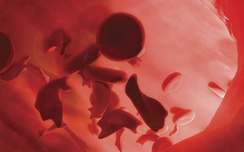
Elevated levels of both types of Lutheran carrier molecules are found on red cells from sickle cell patients. As sickle red cells adhere to α-5-chain containing laminin preparations, it is possible that adherence of sickle red cells to laminin may be relevant to the vaso-occlusion occurring in vivo in patients with sickle cell disease.
Antibodies and their clinical significance
Anti-Lua and anti-Lub tend to be IgM and/or IgG (with anti-Lua being most often IgM). All of the other specificities tend to be found only as IgG (with the possible exception of anti-Lu21, which may contain an element of IgM).
With rare exceptions, and where data is available, Lutheran antibodies cause neither a haemolytic transfusion reaction (HTR), nor haemolytic disease of the foetus and newborn (HDFN). Even where an HTR or HDFN is reported, the symptoms are reported as mild.
In the case of HDFN, this can be explained by the fact that Lutheran antigens are only weakly expressed on cord red cells. In addition, maternal Lutheran antibodies are thought to be adsorbed onto foetal Lutheran carrier molecules expressed on the distal side of the placental tissue, in preference to foetal red cells.
Malcolm Needs was the Reference Service Manager at NHSBT – Tooting Centre and is an IBMS Scientific Advisory Panel member.