For the first time, researchers have shown how the iKnife can accurately detect endometrial cancer before treatment decisions are made.
About 9700 women and people with gynaecological organs are diagnosed with endometrial cancer every year in the UK. The diagnostic process typically involves sending large numbers of people with abnormal, often postmenopausal bleeding, to urgent assessment clinics for investigations. Usually a scan is performed and a biopsy taken for histological diagnosis.
The aim is to rule out endometrial cancer and thankfully more than 90% of people going through the procedure will not receive a diagnosis. But the result of this pathology can take some days and weeks to come back. Everyone awaiting results will likely be very anxious and those with cancer will face delays in treatment.
“We wanted to find a way of making the diagnosis on the biopsy at the point of taking the tissue,” says Professor Sadaf Ghaem-Maghami, Chair of Gynaecological Oncology and Honorary Consultant in Surgical Gynaecological Oncology at Imperial College London, who has led a research team in diagnosing womb cancer using electrosurgical methods.
Diagnostic accuracy
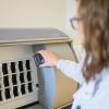
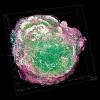
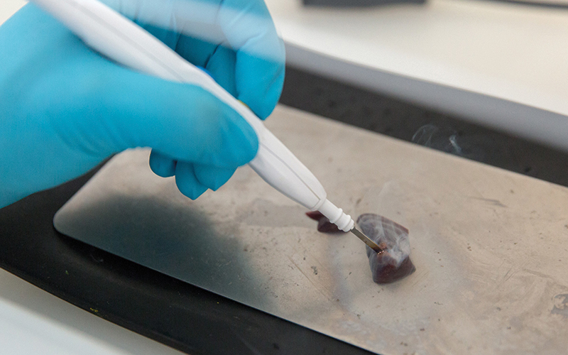
The team used rapid evaporative ionising mass spectrometry (REIMS), the iKnife, to assess biopsy tissue – taken from the uterus using a device called a Pipelle – from 150 women who had endometrial biopsies for abnormal vaginal bleeding. The iKnife works by cutting through tissue while delivering an electrical current, heating as it cuts. It then captures and analyses the vapour that is released to spot any “cancer flags” in the phospholipids in the tissue sample.
“We used the iKnife to burn through the tissue to obtain a mass spectroscopy spectra from the tissues,” Ghaem-Maghami explains. “We constructed a diagnostic model using the REIMS software and the endometrial tissues and compared the results to histopathological routine diagnostic tests.”
“It captures and analyses the vapour that is released to spot any ‘cancer flags’”
The team found the results from the iKnife could differentiate between normal and cancerous womb tissue with a diagnostic accuracy of 89% compared with the “gold standard” for making tissue diagnosis – biopsy. “The sensitivity, specificity, positive predictive value and negative predictive value were 85%, 93%, 94% and 85%, respectively,” Ghaem-Maghami says.
“This means that if the iKnife suggests there is endometrial cancer, we can be quite confident that this is the case, and instigate further tests and start the process of arranging treatment for the patient. We can also reassure women that do not appear to have cancer, so that they do not live with anxiety of their diagnosis for further weeks of uncertainty.”
The iKnife has previously been used for intraoperative diagnosis of resection margins and Ghaem-Maghami and colleagues have also used the instrument for intraoperative assessment of ovarian masses. “However, this is the first time that the instrument has been used to make a diagnosis in the clinic prior to decision-making regarding treatment,” she adds. “To have a technology that may confidently match the performance of histopathology is groundbreaking.”
The accuracy of the technology is “outstanding”. “The amount of tissue obtained from endometrial biopsies is very small and often very scanty and mostly liquid,” Ghaem-Maghami says. “We had to use a second-pass biopsy for the research part of the diagnosis, making the tissue even less adequate. Despite these issues, the iKnife was very accurate in predicting the tissue diagnosis.”
Sadaf Ghaem-Maghami
- 2019 Professor of Gynaecological Oncology at Imperial College Healthcare NHS Trust
- 2015 Began work as private gynaecologist and colposcopist, 132 Harley Street and Consultant Gynaecologist, the Wellington Hospital and Platinum Medical Centre, and as reader in Gynaecological Oncology, Imperial College London
- 2006 Consultant Gynaecologist and Gynaecological Oncologist (honorary), Imperial College Healthcare NHS Trust, and Senior Lecturer, Imperial College London
- 2004 Undertook specialist training in northwest Thames region in obstetrics and gynaecology, which was accredited by the Royal College of Obstetricians and Gynaecologists. Became BSCCP-accredited Colposcopist
- 2002 Honorary Consultant at Queen Charlotte's and Chelsea Hospital, Imperial College NHS Trust
- 1997 Awarded PhD in Immunology
- 1990 Qualified from The London Hospital Medical School (University of London).
Improving outcomes
Like many women in the profession, Ghaem-Maghami juggles work as a clinician, scientist and mother of two girls. “Balancing different demands on my time has on occasions been challenging. Having supportive work and family environments has helped manage these demands, almost successfully.”
She is passionate about women’s health, researching gynaecological cancers for the last 20 years. “It is a real privilege to look after women in the clinic but also be able to advance science, no matter how small the step may be, to provide them with better care and survival outcomes,” she says.
Incidence of endometrial cancer has risen significantly in the recent years and Ghaem-Maghami points to the pressures on the NHS meaning there are more delays in diagnosis for women with abnormal bleeding. “I wanted to be able to change the way these women experience care and help improve their outcomes. If endometrial cancer is diagnosed early, it is cured in the majority of cases, so it is essential that we aim for a diagnosis as quickly as it is accurately possible,” she adds.
The research team is planning a large clinical trial in multiple centres across the UK and Europe to assess the diagnostic accuracy of the technology in a “real world” setting in different centres. The hope is that the technology will be integrated in clinical care pathways and will accelerate diagnosis of endometrial cancer.
“It is too soon to say that we will not need pathological diagnosis, but to be able to make a fairly accurate diagnosis at the point of taking the biopsy will have a significant impact on clinical practice and the way we treat these women,” she adds. The result should be earlier diagnosis and expedited treatment of endometrial cancer, leading to better outcomes and better patient experience.
Image credit | Thomas Angus_Imperial college London