Senior Biomedical Scientist Alix Costello with a nuts-and-bolts guide to creating a new histology laboratory, with insights into some of the new and exciting apparatus now available for modernised working practices.
My name is Alix Costello. I recently obtained a post at Blackpool Victoria Hospital as a Senior Biomedical Scientist following completion of the Scientist Training Programme in Histopathology. One of my first roles after starting in the department was to help coordinate a laboratory move while maintaining our diagnostic services. Yet, to get to this stage, a lot of hard work had been going on behind the scenes to facilitate the procurement of the new histology laboratory within just fourteen months. This article will provide an insight into the planning and design process of building a new laboratory and explore some of the bespoke equipment pieces designed to modernise our histology practices.
Why and how
In April 2021, a decision was made that the 50-year-old, tired histology estate was no longer a suitable workspace for the histology team. A project began to design a new, modernised laboratory in a disused space adjacent to the existing histology laboratory. Once the decision had been made to move forwards with the plans for the new laboratory, it was clear that to meet and futureproof the needs of the department, specialist design consultation would be necessary. This was important to not only maximise the physical design of the laboratory workspace, but to ensure that the services installed would help safeguard staff, not only to current standards but also to future evolving regulations for containment and working practices.
A specialist design consultancy was appointed and tasked with designing the facility but they worked closely with the histology team to ensure that design concepts would be technically and practically feasible. Utilising specialist knowledge and experience from the histology team, the design consultancy worked to create an open-plan workspace that would facilitate lean working practices and maximise the estate’s potential. Following the design process, the tendered specialists in laboratory equipment finalised designs for bespoke histology equipment with significant focus on local exhaust ventilation (LEV) to safeguard staff, not only to current standards but also to future evolving regulations.
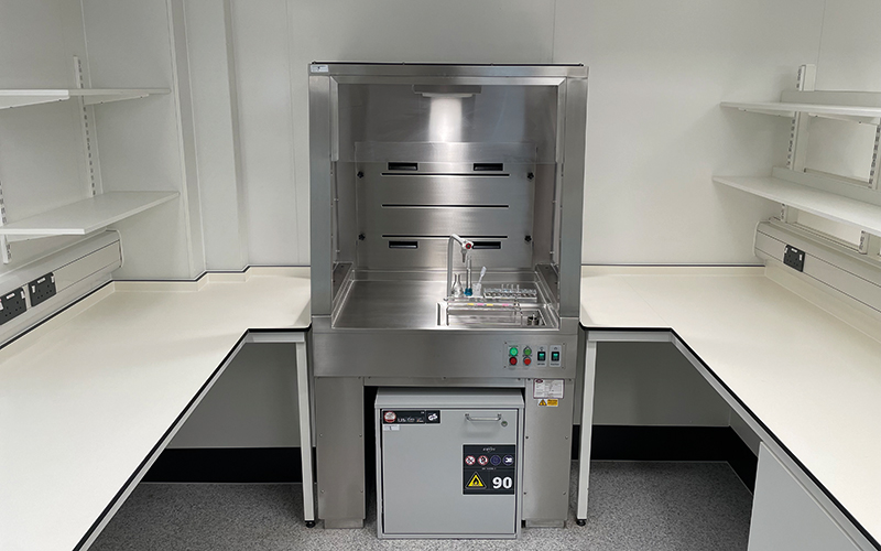
In histology, technicians commonly work in close proximity to hazardous reagents, most notably formalin, xylene and alcohol. Despite the dangers associated with using these reagents, (particularly formalin, which is a carcinogen, mutagen and sensitiser), they are essential to histology practice. It was therefore vital that a strong element of the new laboratory design process and focus for the new equipment was resilience for both established and future health and safety practices. With this in mind, several new, built-in pieces of equipment were commissioned, which transformed and modernised our working practices whilst maximising safety for the histology staff.

Bespoke equipment
1. Cut-up space: The old histology laboratory had one bench dedicated to surgical dissection work and one bench for handling biopsy specimens. During busy periods this led to bottlenecks as the workflow was restricted by the spatial constraints. In the new laboratory, we now have three benches available for surgical cut-up and one dedicated to biopsy specimens. Each of the benches is height adjustable to maximise user safety.
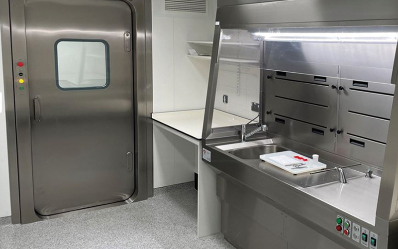
3. Containment Level 3 (CL3) room: Blackpool Victoria Hospital is a cardiothoracic hospital so the laboratory receives a high volume of lung frozen section work to facilitate intra-operative diagnosis. In the old laboratory, our cryostat was located in the very cramped cut-up room. Additionally, having just one cut-up bench meant that surgical dissection work had to pause to enable frozen sections to be performed on the ventilated work bench. Working with fresh lung tissue carries an infection risk, therefore the new laboratory project seized the opportunity to build a CL3 room for handling frozen-section work. The CL3 room can be used for handling infectious tissue specimens and acts as a spare cut-up bench.
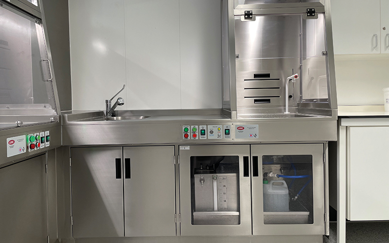
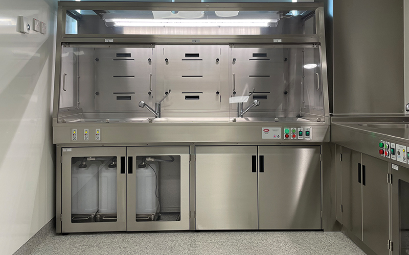
6. Staining facilities: We also now have LEV units surrounding our special stains workbench, routine staining and coverslipping equipment. This has further improved safety for our staff and reduces exposure to hazardous reagents.
Clean room: Finally, as Blackpool Victoria Hospital is a cardiothoracic centre, we receive lung biopsies and resections that require molecular pathology investigations. In 2018, we started a molecular pathology service and we now provide this service for lung cancer patients across all four trusts in the Lancashire region. As part of our service, we are able to test tumour tissue for mutations to guide patient treatment. The technique that we use utilises real-time PCR so it must be performed in a clean working environment to reduce risk of cross-contamination and aberrant results. In the old histology laboratory, the molecular pathology investigations had their own “clean” microtome but otherwise preparations took place in the same room as immunohistochemistry due to spatial constraints. As part of the new laboratory project, a separate “clean” room was built into the immuno-histochemistry workspace to enable sample preparation for PCR testing to be performed separately.
Move-in day
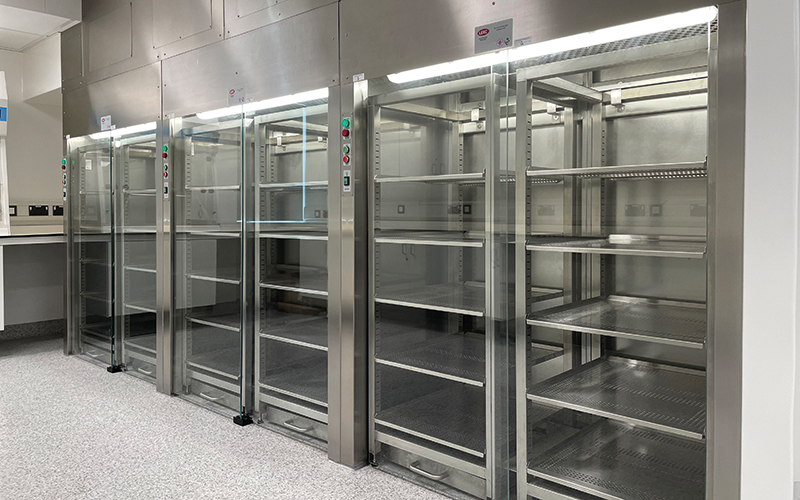
In June 2022, after 14 months of planning and building, the new histology laboratory was ready for use. While everyone was excited and raring to get started in the new workspace, the laboratory move needed to be planned carefully to prevent disruption to the service for our users. Suppliers and engineers were scheduled for equipment moves, which occurred gradually over a two-week period. The old laboratory and the new laboratory were adjacent to each other, so we were able to work across both spaces for a couple of weeks while the move took place. We also wanted to use this opportunity to adapt our laboratory workflow to maximise workspace and embed lean working practices. By the end of June, we had completely moved into our new laboratory and had started to get to grips with new ways of working.
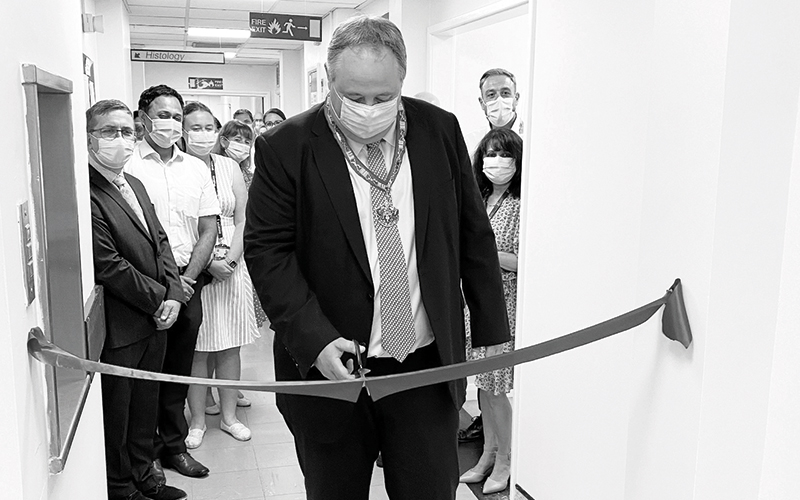
Laboratory opening
Later in the summer, we had our official laboratory opening, including the cutting of a red ribbon, The President of the Royal College of Pathologists Professor Michael Osborn travelled to Blackpool to officially open the new laboratory. I also marked the official opening by creating a piece of artwork, which is now hanging by the front door of the new laboratory.
Benefits of the new laboratory:
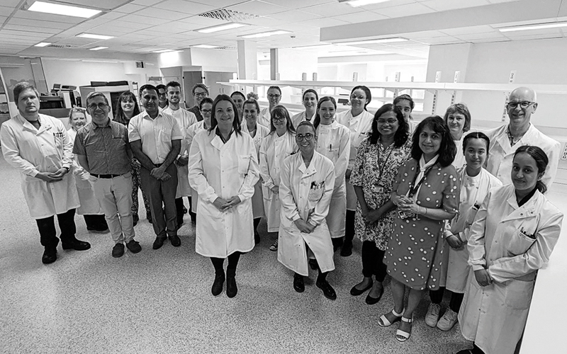
Six months later, I can definitely say that the entire histology team feels very fortunate to be working in this new environment. The new laboratory has brought about a number of benefits:
- An open-plan workspace that has enhanced team communication
- A fresher and brighter laboratory that has improved staff working conditions
- Greater space for storage and equipment has meant that individuals can perform their roles more efficiently
- Increased cut-up bench capacity has significantly reduced bottlenecks within the department, helping to improve diagnostic turnaround times
- Brand new LEV systems throughout the laboratory have enhanced safe working practices and futureproofed the department against evolving safety standards
- Increased bench space has facilitated an increase in desktop computers, which will reduce reliance on paper documentation.
Conclusion
In summary, the 14-month long project has already had significant benefits for laboratory staff and service users. Our previous laboratory was built to suit cellular pathology practices from the mid-1900s, practices that are quite unrecognisable from today’s automated machinery and growing, increasingly complex workload.
We recognise that we are very lucky to now work in a purpose-built facility with bespoke safety mechanisms that complement our working practices. However, we realise that for the majority of histology laboratories this is not a reality. By writing this article we aim to provide an insight into some of the new and exciting histology apparatus now available for modernised working practices. We would be happy to share our learning from the project with other histology teams going through a similar process.
Alix Costello is a Senior Biomedical Scientist and Clinical Scientist at Blackpool Victoria Hospital
Image credit | iStock | Blackpool-Cellular-Pathology-Laboratory