The IBMS has awarded five research grants this year. Here, the first two successful candidates outline the work they are undertaking.
José Ignacio Saldaña
Lecturer in Immunology, University of East London
Antibody crosslinking of the cell surface receptor Siglec-F: a method to study alveolar macrophage function and adaptation
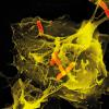
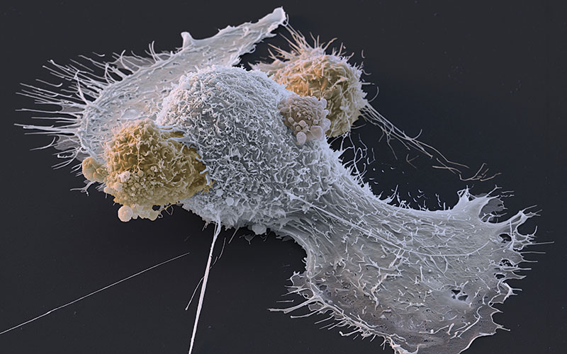
Macrophages are highly diverse immune cells that carry out functions that are specialised to the characteristics of the tissue in which they exist.
As these cells move in, they experience a process of adaptation driven by signals provided by local factors and by the physiological environment. Therefore, macrophages of the airways (alveolar macrophages), liver (Kupffer cells) and brain (microglia) have very different roles and can be differentiated from one another.
Lung tissue is delicate and constantly exposed to particles, dust and pathogens contained in the air. Upon detection of pathogens, alveolar macrophages are quick to respond to protect the airways, being highly pro-inflammatory. These cells are kept under a regulated state thanks to close contact with epithelial cells that provide negative controlling signals through specific cell surface proteins. This regulatory communication system is essential, as any unwanted inflammation can damage the delicate lung tissue or induce scarring and remodelling, leading to loss of function.
Other proteins (e.g. mucins and collectins) are ubiquitous components of the extracellular matrix of the lung that also deliver developmental and regulatory signals to alveolar macrophages through receptors such as Siglec-8 and its mouse counterpart Siglec-F. The specific role of Siglec-F, as a regulator of the process of macrophage function and adaptation to tissue microenvironments, is not fully understood. Therefore, targeting and modulating signalling through Siglecs has the potential to create new specialised therapies.
Dr Saldaña’s IBMS Research Grant-funded project aims to target Siglec-F and uncover the potential effects this could have on resident lung macrophages, function and tissue adaptation.
Thomas McDonnell
Research Associate, University College London
Common pathways in systemic lupus erythematosus and antiphospholipid syndrome
Systemic lupus erythematosus (SLE) and antiphospholipid syndrome (APS) are autoimmune disorders characterised by autoantibodies, activation of inflammatory and coagulation pathways and an adverse impact on quality of life. Characterised by thrombosis, strokes and/or recurrent pregnancy loss caused by antiphospholipid antibodies (aPL), APS may occur alone or in combination with SLE. Systemic lupus erythematosus is a multisystem disease with potentially serious organ and life-threatening manifestations in which anti-dsDNA antibodies fluctuate with disease activity. Despite the clinical severity of these diseases, treatments are imperfect and lack specificity against autoantibodies.
Approximately 40% of APS and SLE patients develop antibodies to serine proteases, principally factor Xa (FXa) and/or thrombin (Thr). The relevance of these antibodies to disease is currently unknown. As both FXa and Thr are therapeutic targets in other disorders and coordinate interactions between complement and coagulation cascades, it is tempting to speculate that antibodies directed against these factors may be important in SLE and APS.
Both the coagulation and complement pathways are proteolytic cascades of serine proteases and are activated in both diseases. In SLE, complement consumption is a biomarker of disease flares, and low C3 levels are associated with active disease. In APS, complement activation is associated with pregnancy morbidity and the generation of thrombosis in mouse models.
Interactions between coagulation and complement cascades are poorly characterised. FXa and Thr have been shown to cleave complement factor 3 (C3) and 5 (C5) that are central to complement activation; however, the precise function of antibodies to FXa and Thr are unknown and Dr McDonnell hypothesises that they promote complement activation in patients with SLE and/or APS.
After consultation with patients, 88% to 90% of 527 respondents felt this work was important and relevant to management of these diseases, confirming patient/public interest in this mechanistic research evaluating the impact of autoantibodies on FXa-and Thr-mediated complement cleavage in SLE and APS.
To determine whether or not these antibodies are associated with complement cleavage in patients, Dr McDonnell looked for clinical correlations between anti-FXa and/or anti-Thr IgG positivity and C3 levels in patients with SLE under long-term follow-up at University College London Hospital. Patients with either anti-FXa and/or anti-Thr IgG positivity were identified and their C3 levels examined from five previous consecutive clinic visits. Significantly, lower levels of C3 were found in patients with both anti-FXa and anti-Thr IgG positivity than with single anti-FXa or anti-Thr IgG positivity.
Dr McDonnell’s research has focused on the cleavage of C3. His research grant-funded further study will examine the effects of anti-FXa and anti-Thr antibodies upon FXa- and Thr-mediated C5 cleavage, which is important in the formation of the membrane attack complex that mediates cell lysis. The aims are i) to assess anti-FXa/Thr activity on complement cleavage and AT-mediated inhibition; and ii) develop a system applicable to serine protease-mediated complement generation and cleavage in endothelial cells.
Image Credit | Science photo library