Yvonne Webb, a Specialist Biomedical Scientist from NHS Greater Glasgow and Clyde, looks back over a case study.
The patient was a 37-year-old female with a history of pre-eclampsia, HELLP syndrome (which is the acronym for “haemolysis, elevated liver enzymes, low platelets”) and a medical termination of pregnancy at 23 weeks the previous year. At the booking visit, the patient grouped as an A Negative, antibody screen negative.
Three months later, the patient arrived in the Maternity Assessment Unit with pre-eclampsia and was admitted for monitoring. This is a condition that can develop from 20 weeks in pregnancy. It is usually identified by a high blood pressure measurement in women who have previously not experienced high blood pressure. They will have a high level of protein in their urine and often swelling in the feet, legs, and hands.
HELLP syndrome is a life-threatening liver disorder that is thought to be a type of severe pre-eclampsia. It is characterised by haemolysis, high levels of liver enzyme, which can indicate liver damage, and a low platelet count. The only way to treat severe pre-eclampsia and HELLP syndrome is to deliver the baby.
The ward had sent down a Group and Save sample. The initial three-cell antibody screen was positive: 1+ in screen cell 2. Antibody identification panels were also done.
The results
Anti-D by enzyme and inconclusive by IAT (not present in screen cell 1 and panel cell 2).
The patient’s phenotype was ccdee K- and the titre was 1/1. The midwife looking after the patient had confirmed she had not had any prophylactic anti-D this pregnancy. A sample was requested from the partner and two samples sent to Scottish National Blood Transfusion Service (SNBTS) for quantification. The partner sample was tested and found to be Group O RhD Neg, phenotype ccdee K-.
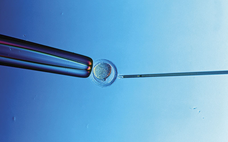
I carried out the testing in the early hours of the morning, so double-checked all my work. After a discussion with the midwife, she double-checked with the patient again that she hadn’t had any anti-D and was concerned about how this was even a possibility. The midwife and the medical registrar investigated further and discovered that the patient was undergoing an IVF pregnancy and was a recipient donor egg.
Post-analysis
There was miscommunication by an inexperienced biomedical scientist who was on shift that day, as they had spoken to the SNBTS consultant. As per British Society for Haematology guidelines, and in the absence of SNBTS being able to determine whether the anti-D was immune or prophylactic, anti-D was issued.
Verbal results from SNBTS
No red cell antibodies present by IAT. Irregular results present in enzyme – possible weak anti-D specificity.
This difference was due to SNBTS using different analysers (ORTHO used in SNBTS, BIORAD used in our lab) and manufacturers of antibody identification panel cells.
Later that night, the biomedical scientist passed all these details on to me. I had discussed with them that the patient had not had any prophylaxis anti-D and, therefore, had to be immune if they have proceeded to look at patient notes in our LIMS system. I had a conversation with our haematology consultant and had agreed that the patient did not require any prophylaxis. However, the patient had already been given the anti-D authorised by the SNBTS consultant earlier that day. The patient’s pre-eclampsia was getting worse and the consultant decided to deliver baby at 28 weeks, rather than sending sample for cffDNA genotyping. The patient had a unit of A Rh Neg blood (rr, K-) following her caesarean section.
Prophylactic anti-D was given and we are unable to determine if the patient’s own immune anti-D was still present. After further bleeding post-caesarean section, the patient had another two units given A Rh Neg (rr, K-).
An antibody investigation indicated the possibility of a new antibody forming.
Was this from the units transfused or from the baby?
A Kleihauer was carried out after delivery and the patient was found to have 5.1ml bleed. The SNBTS consultant advised to give anti-D. The baby’s group was analysed urgently– blood group: O Rh Pos. Phenotype: CcDee
I spoke briefly and exchanged a few emails with the patients’ consultant, who confirmed that the patient hadn’t been aware that the egg was RhD positive and it wasn’t stated in her pregnancy management notes. The consultant had stated foetal free DNA typing (cffDNA) wouldn’t be required as the IVF clinic had told the patient the egg was RhD positive when she contacted them after the results of her antibody screen and her partner testing results.
Unanswered questions
How did the IVF clinic know the egg was Rh Pos? What were their guidelines and did they adhere to them? Would the IVF clinic give Rh Neg woman a Rh Neg egg?
Mandatory IVF requirements
The donors must be negative for HIV1 and 2, HCV, HBV and syphilis on a serum or plasma sample tested as follows, namely:
- HIV1 and 2: Anti-HIV – 1, 2
- Hepatitis B: HBsAg and Anti-HBc
- Hepatitis C: Anti-HCV-Ab In certain circumstances, additional testing may be required depending on the donor’s history and the characteristics of the gametes donated (for example, RhD, Malaria, T.cruzi).
I had briefly spoken to a Quality Manager from an IVF clinic about the mandatory requirements and the response was that they don’t screen and consider Rh of the donor egg unless the patient has antibodies. So my question to that was: “Why did the clinic tell the patient they knew her egg was Rh Pos if they don’t routinely test for it?” The Quality Manager couldn’t answer that question.
Follow up
The laboratory haematology consultant has written to the obstetrician looking after the patient to have a repeat sample done six-months post-delivery to try to confirm whether the antibody is immune or not and to identify the possibility of any new antibodies which were developing following the delivery and multiple transfusions.
Image Credit | Science Photo Libary