Andrea Johnson, Cellular Pathology Laboratory Manager, gives a tour of her lab in Herefordshire.

I am the Cellular Pathology Laboratory Manager at Wye Valley NHS Trust, a small laboratory in rural Herefordshire, processing 21,000 histology and 1300 cytology specimens a year. I enjoy networking, especially having the opportunity to talk to others in the discipline. Several people have said to me recently that there is a lot to be learnt from smaller laboratories. Why is this? It’s because we have had to adapt and evolve.
We have a dedicated, patient-focused team who work flexibly to ensure patient specimens are treated on an individual basis. We have developed the range of work our medical laboratory assistants (MLAs) complete and our microtomes are usually manned by our MLA team. Our biomedical scientist team has experienced rapid expansion recently, allowing the team to take on numerous IBMS qualifications and develop advanced biomedical scientist dissection, with biomedical scientists now completing around 99% of all specimen dissection.
We are also supported by a dedicated admin team who work incredibly hard with multidisciplinary team preparation, molecular referred work and specimen tracking.
We are an incredibly efficient laboratory with no specimen or block backlog.
I believe many of our efficiencies stem from our managed laboratory service (MLS) and of course teamwork. Our MLS provider supplied an excellent laboratory tracking system, which is linked to our laboratory information management system (LIMS). The tracking system implementation eight years ago allowed us to significantly reduce paper records, and ensure audit trails and traceability for all blocks and slides. The system allows us to print slide labels at microtomy that can be placed straight on to the immunohistochemistry (IHC) stainer or digital pathology scanner.
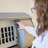
Wye Valley NHS Trust was the first hospital in the West Midlands Cancer Alliance initiative, across four NHS pathology networks, to go live with digital pathology on 1 November 2022. We are currently in the process of clinical validation and are excited to be installing a second scanner in April this year. We are also in the process of a number of new developments that aim to increase our test repertoire and ensure we can work to maintain the highest level of patient care. This year will see us add an additional IHC stainer, dissection imaging system and automated temperature and fume monitoring.
While the laboratory boasts technology, efficiency and no backlog, we do have a very significant issue. We are struggling to recruit histopathologists, which is understandable given the national shortage. Our progress with digital pathology and the dissection imaging system is a significant opportunity to recruit pathologists for digital reporting with the flexibility of home working.
We, like all pathology laboratories across the country, are involved in the development of a pathology network, and are a member of South Midlands Pathology (SMP). Networking provides exciting opportunities to work collaboratively to ensure the right result for the right patient at the right time. Looking at patient pathways and harmonising practices, along with the implementation of digital pathology and the future of a single
LIMS, allows us to adapt and overcome our reporting shortage with a network-wide approach.