A guided tour of the Oxford Autoimmune Neurology Diagnostic Laboratory.

Autoantibodies against proteins expressed in the nervous system cause disease. The discovery was made at the turn of the century, since which a group of antibody-mediated, often treatable, diseases have been defined based on the presence of target-specific antibodies in the serum or cerebrospinal fluid in people who present to neurology, ophthalmology or psychiatry. Our diagnostic lab in Oxford has emerged from our role in the discovery of some these antibody targets.
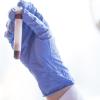
Circulating antibodies can only bind to targets accessible in the extracellular space – either circulating soluble molecules, molecules bound to receptors on the extracellular surface, or the extracellular portion of membrane proteins. Assays that closely replicate these surfaces appear the most accurate for autoantibody detection and patient care. We run about 30,000 live cell-based tests a year where integral membrane or soluble proteins are overexpressed in living cells.
We run tests for people who present with different neurological issues, such as inflammation of the optic nerve or optic chiasma, the spinal cord, or brain, those who develop seizures, stiff person or stiff limb syndrome, or progressive encephalomyelitis with rigidity and myoclonus.
Autoantibodies also cause diseases of the peripheral nervous system, such as autoimmune myasthenia gravis (MG), in which autoantibodies to the nicotinic acetylcholine receptor fix complement and damage the neuromuscular junction, causing fatigable muscle weakness. AChR antibodies are routinely detected by radioimmunoassay in immunology labs. However, the ability to cluster AChR with the cytosolic molecule RAPSYN in live cell-based assays improves test sensitivity and identifies people, negative by AChR RIA, who have AChR autoantibodies but were considered to have seronegative MG. This distinction is important, particularly with new therapeutics that target the complement system. A second target, muscle-specific kinase, discovered in part by past members of our team, is run alongside the adult and fetal AChR as a panel.
The final group of autoimmune neurological diseases were first described 10 years ago. Now termed autoimmune nodopathies, these are defined by the presence of antibodies to nodal or paranodal proteins (neurofascin-155 or -186, contactin-1 or CASPR1). They cause damage to peripheral motor or sensory nerves, resulting in disabling paralysis, unsteadiness, and tremor, and can sometimes impair swallowing or breathing. The usual treatments used for seronegative inflammatory neuropathies are often ineffective in the presence of these antibodies, and alternative approaches are required.
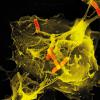
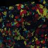
For people with a high suspicion of a nervous system antibody-mediated disease with no known target, assays using primary cultures of live neurons, human stem-cell derived peripheral neurons and myelinating co-cultures, astrocytes or indirect immunohistochemistry on rodent brain sections may identify an antibody. Additionally, we run other assays for rare targets, such as the glycine receptor and ganglionic AChR, amongst others. The methods that we use are the most accurate way to detect neurological antibodies.
Funding from the Sumaira Foundation and the Africa Oxford Initiative has supported the development of a training programme for scientists and neurologists from low- and middle-income countries to visit us in Oxford to learn the live cell-based assay technique. Guests from Africa, Asia and the Middle East have each spent four weeks in our laboratory and we are now working with the Sumaira Foundation to establish these tests in Iran, Sri Lanka and Zambia.
Lab facts
We run about 30,000 live cell-based tests a year, where integral membrane or soluble proteins are overexpressed.
We run tests for people who present with issues of the central and peripheral nervous systems. They can present with disturbances in their visual or motor skill, impaired breathing or swallowing, or bladder and bowel disability.
Funding has supported a training programme for scientists and neurologists from low- and middle-income countries to visit us and learn live cell-based assay technique.