New research has led to the UK government approving the use of digital pathology to help speed up analysis of cancer screening samples.
This allows the benefits offered by digital pathology to be used to improve cancer screening, particularly in bowel, breast, lung and cervical cancers.
The use of this technology will result in faster reporting of people’s samples.
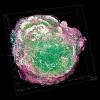
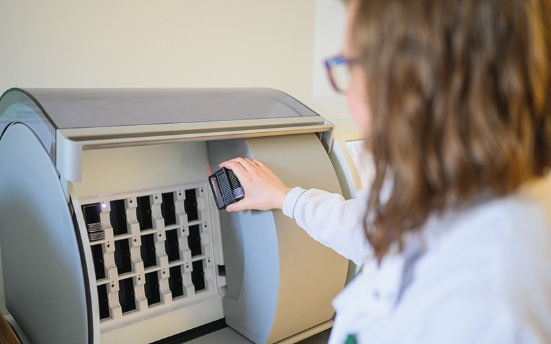
Digital pathology is the use of automated slide scanners to digitise the histopathology process, with results reported on computer workstations, as opposed to a conventional microscope, enabling pathologists to report samples remote from the laboratory producing slides.
This process makes sharing samples easier, helping to reduce risk of loss or damage of samples.
It also mitigates the need for pathologists to be present in hospitals, as they can review the slides remotely. Digitising the slides might also allow the use of computer algorithms to help improve pathologists’ performance in the coming years.
The research was funded by the National Institute for Health and Care Research and the news comes after a consultation that was conducted by the UK National Screening Committee.
Lead Researcher Consultant Pathologist Professor David Snead said: “I am delighted that digital pathology is cleared for use in cancer screening programmes.
“It is a big milestone to achieve and we are extremely proud that the work we have led proved so effective in making this change.”
Image credit | University of Warwick