Research has found that cellular structures act as “molecular plasters” that protect against infections, such as tuberculosis. We talk to study co-author Claudio Bussi.
When laboratory work was severely restricted during the COVID-19 pandemic, Dr Claudio Bussi, Postdoctoral Fellow at the Francis Crick Institute, had more time for catching up on literature. “At that time, many papers in cell biology began to explore the concept of membraneless organelles and phase separation in more detail,” he says.
“The idea of an organelle without a physical membrane was very exciting for me and piqued my curiosity. I was nearing the end of my first postdoctoral project and thought I could develop a second project exploring these concepts.”
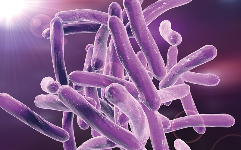
Claudio searched for transcripts associated with stress granules (SGs) – membraneless organelles that appear in response to stress in the cytoplasm – within an old RNA-seq dataset generated by his lab, which studies host–pathogen interactions in Mycobacterium tuberculosis (Mtb). “I chose SG because it is one of the best-characterised membraneless organelles with defined markers for investigation,” he adds.
He found that many SG-associated transcripts were upregulated during Mtb infection, but this phenomenon was not observed during infection with an Mtb mutant strain that has a restricted ability to cause membrane damage. “This was certainly a promising sign, so the next obvious step for me was to try to visualise the process and see if I would observe a phenotype worth further investigation,” says Claudio.
Repairing cell damage
What followed was a study that uncovered a biological function for the SG. “When I finally regained access to the microscope to check if this response was triggered under conditions that cause membrane damage, such as Mtb infection, I was fascinated by how clear and strong the phenotype was.
I anticipated an increase in SG formation after inducing damage, but I was not expecting such a robust response spatially linked to it.”
“Observing the rapid formation of granules after inducing lysosomal damage was quite exciting”
During infection with Mtb, immune cells called macrophages are recruited to envelop the bacteria and remove the threat. But the bacteria can secrete chemicals to damage the macrophage phagosome membrane – where the bacterium resides after being internalised by the host – and escape their “immune prison”. When macrophage membranes are ruptured, SGs rapidly form a plaque to plug the gaps, allowing for cellular repair machinery to come and fix the damage.
“Observing the rapid formation of granules after inducing lysosomal damage and their close association with lysosomes was quite exciting,” says Claudio. The team also showed that the ability to recruit these “molecular plasters” was essential to keep infection under control. When they edited infected cells to remove genes responsible for SG formation, macrophages could no longer envelop and destroy bacteria, allowing the infection to take over.
“The most thrilling part came when a good friend of mine – Agustín Mangiarotti, a postdoc at Max Planck Institute of Colloids and Interfaces in Germany – who serendipitously turned into a collaborator in this project, managed to reproduce these findings in vitro,” Claudio says. “The ability to literally visualise a molecular plug being formed specifically at the site of damage to repair a vesicle was very surprising.”
Challenges
The findings were confirmed again in silico by Christian Vanhille Campos, a PhD student at the Institute of Science and Technology in Austria. Computer models of organelle damage and repair showed similar patterns of plaque formation over areas of damage.
“Finally, when we obtained gene-edited cells that were unable to form these granules and observed that they were almost incapable of recovering after inducing damage, it provided definitive confirmation that we were reporting a very important cellular function,” Claudio says. “We were all very excited about this discovery.”
The research was not without its challenges, one of which was working with induced pluripotent stem cell (iPSC)-derived macrophages, which are not as easy to handle as commonly used cell lines. “While these cells offer the significant advantage of being primary-like and, hence, more physiologically relevant, certain applications, such as routine maintenance and live-cell imaging, pose greater technical challenges,” Claudio says.
Claudio Bussi

- Postdoctoral Training Fellow – Host–pathogen interactions in tuberculosis at the Francis Crick Institute, London
- Received a PhD in cell biology – Innate Immunity, Universidad Nacional de Córdoba in Argentina
- Undertook PhD Internship, cell biology – Live-cell imaging, The Babraham Institute in Cambridge.
Future research
The findings have implications for addressing new therapeutic strategies in infections where membrane damage occurs, such as TB, but other diseases where cell and organelle damage can take place, like cancer and neurodegenerative diseases, such as Parkinson’s disease.
The team plans to continue studying the role of SGs, and possibly other similar molecules, to find ways of boosting natural defences against infection. “I believe the field exploring the interactions of different biomolecular condensates with membrane-bound organelles is in its early stages,” Claudio says.
“There is much more to explore in this area, including determining what regulates these contacts and the contexts in which they occur. Considering that many proteins relevant to a wide range of diseases accumulate in these condensates, I believe this field holds promising therapeutic potential as well,” he adds.
Claudio has been inspired to focus on cell biology in his career and has a passion for “intracellular life” and microscopy, especially live-cell imaging. “The ability to develop tools that visualise and validate biological functions is incredibly fascinating. When you witness dynamic processes in real-time, it’s hard not to be curious – you start asking how it’s happening, what regulates these processes, and what their functions are,” he says.
“In my view, just as certain transcriptional programmes define cellular functions, the dynamics of specific organelle networks contribute to cellular identity,” he adds.
His career so far has not been without its issues, and completing his PhD in Argentina, where funding is inconsistent, has added an “extra layer of challenge”. Claudio says: “Over the years, you learn to adapt to situations that consistently place you at a disadvantage on the global scientific stage. Scientists there exhibit remarkable resilience, and I’ve drawn inspiration from them to overcome these challenges.”
These lessons have paid off and the recognition for his recent research is “gratifying and humbling”. Claudio concludes: “There’s still much more to explore in this field, and I’m excited about the possibilities.”
Image credit | Science-Photo-Library| iStock