In 2019, the IBMS awarded research grants totalling almost £27,000. Here, the seven recipients explain their grant-supported projects.

Head of the Betsi Cadwaladr University Health Board North Wales Clinical Research Centre
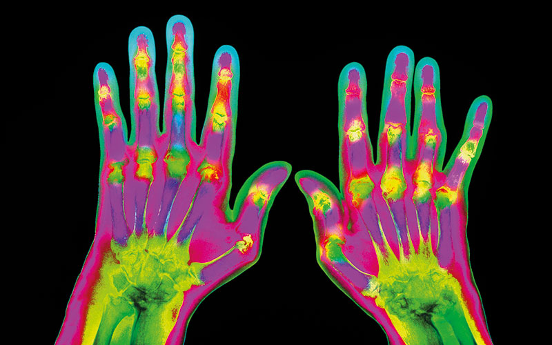
Pilot study of adjunctive therapy with Losartan and Allopurinol in Patients with Rheumatoid Arthritis and Interstitial Lung Disease
The North Wales Clinical Research Centre (NWCRC), based at Wrexham, led by Professor Stephen Fôn Hughes, initiative by the Betsi Cadwaladr University Health Board (BCUHB) Research and Development Department, was designed to encourage and support research within the health board and to promote clinical research across North Wales and beyond.
The proposed study has provided an exciting opportunity to work with Professor Mark Garton, a consultant rheumatologist, based at Wrexham Maelor Hospital, North Wales.
Rheumatoid arthritis (RA) carries a 10% lifetime risk of interstitial lung disease (RA-ILD), with median survival of two to three years. RA-ILD causes fibrosis (thickening) of lung tissue, variable inflammation and impaired gas exchange. Patients report dry cough/exertional breathlessness, often refractory to (or aggravated by) conventional therapies.
RA also increases cardiovascular risk, reflecting systemic inflammation, higher rates of traditional cardiovascular risk factors, and side-effects of corticosteroids and non-steroidal anti-inflammatory drugs.
Due to the lack of novel studies in this area, this research promises to increase subject knowledge in this field. Patients with RA-ILD will benefit from better ascertainment of early ILD and targeted medication, which should definitely reduce vascular morbidity while possibly slowing ILD progression.
Senior Lecturer in Biochemistry, University of Worcester
Markers for Alternative Treatment Strategies for Acute Myeloid Leukaemia
This work is about understanding how heamatopoietic stem cells develop and on determining the biological events which occur when stem cell development goes wrong, causing leukaemia. This project focusses on acute myeloid leukaemia (AML) – the most common acute blood cancer in adults. The disease is genetically very heterogeneous and prognosis varies according to particular genetic abnormalities.
The standard treatment for AML has evolved little in the last four decades and many patients relapse following therapy.
The aim of this study is to determine whether inhibition of the Hedgehog signalling pathway is a viable treatment strategy for different forms of AML. The hedgehog signalling pathway drives cell proliferation and its components are well established as targets for anti-cancer therapeutics. Leukaemic cell lines will be screened for hedgehog signalling markers and monitoring cell proliferation and apoptosis in cells treated with hedgehog pathway inhibitors to determine whether this treatment strategy is applicable to some or all forms of AML.
One negative prognostic marker for AML is expression of myeloid leukaemia factor 1 (MLF1). Since little is known about the protein, it is not a primary choice of drug target. However, research indicates that there is cross-talk between MLF1 and the tumour suppressor protein SUFU, a negative regulator of the hedgehog signalling pathway. This pathway, which is highly active during embryogenesis, is largely quiescent in healthy adult cells and its abnormal activity is associated with many different forms of cancer. In brief, the pathway is activated by binding of the hedgehog ligand to a receptor, Patched, which then relieves its inhibition of another transmembrane receptor, Smoothened. This results in the release of the GLI transcription factor by the tumour suppressor SUFU leading to upregulation of transcription of genes involved in cell proliferation differentiation and survival.
Components of the hedgehog signalling pathway are well-established as targets for anti-cancer therapeutics. Recent in vitro and in vivo studies have demonstrated that targeting hedgehog signalling may be an effective anti-AML strategy including in cases of drug-resistant AML. However, undesirable side-effects have been associated with hedgehog pathway inhibitors.
The aim of the study is to determine the relationship between MLF1 expression and hedgehog pathway activity in leukaemic cell lines, to determine whether MLF1 screening provides a rationale for selective use of hedgehog pathway inhibitors in AML patients.

PhD student, North Wales Clinical Research Centre
The role of routine and novel biomarkers and their correlation with clinical outcome measures in patients undergoing surgical interventions for benign and malignant disorders of the prostate.
The management of prostate disorders presents an increasing demand on already strained healthcare systems in many countries. Although safety for patients undergoing urological surgical procedures is improving, development of post-operative complications remains a serious concern. The aim of this study is to investigate the changes in selected biomarkers after prostate surgeries to test the hypothesis that post-operative changes in biomarkers are associated with the development of post-operative complications. This innovative research will provide additional knowledge to an area that has not been well established and provide predictive markers for post-operative complications.
Findings from this study will provide a single or a panel of blood/urine tests to predict the risk of patients developing post-operative complications. Specific changes to their management could then be instituted to improve patients’ outcomes. This intervention will be beneficial for patients, through timely recognition and management, as well as offer benefits to healthcare providers by reducing treatment costs and readmissions due to post-operative complications.

Professor at the School of Medicine, Faculty of Health & Medical Science, Taylors University, Dr Soo Ching Lee the University of Malaya’s Department of Parasitology.
The microbiome diversity of the Malaysian long house of Borneo and inflammatory bowel disease
The mammalian gut houses trillions of micro-organisms and there is evidence that exposure to a wide range of microbiota throughout life is essential for the development of a competent immune system.
It has been proposed that the incidence of inflammatory bowel disease (IBD) is linked to a lack of biodiversity in modern urbanised environments. An Asia–Pacific study group recently reported on the increasing incidence of IBD in developing Asian countries, to include Malaysia. Consequently, the changing environment has a profound effect on the incidence of both microbiome diversity and diseases of the post-modern era such as IBD.
Malaysia comprises the Malay Peninsula (West Malaysia), and the states of Sarawak and Sabah (East Malaysia), on the island of Borneo. West Malaysia houses the capital city of Kuala Lumpur and has the greatest level of urbanisation. In contrast, much of Borneo comprises natural rain forest housing an abundant biodiversity which greatly influences the natural eco-systems, and which in turn shapes the lives of its people. Sarawak has a population of less than 3 million, with the majority Dayak indigenous communities accounting for 40% of the population. Traditionally, the Dayaks have lived in long houses in the rain forests. Whilst modern day long houses are sometimes comprised of modern building materials, the basic communal concept is retained comprising a long continuous corridor sometimes up to 200 metres long from which individual family compartments shoot.
This research hypothesises that we will see significant differences in the balance of symbiotic and potentially pathogenic microbiota, when comparing the profiles of residents of the rural long house, urban Kuala Lumpur and patients with IBD. With the incidence of IBD rising in the region, it is important to define microbiome profiles of patients with IBD, while at the same identifying elements of the rural microbiome that may protect against the disease.

Course Director for the BSc Biomedical Science, University of Wolverhampton
Assessing a novel CRISPR/Cas9 mouse line as a useful tool for autosomal recessive polycystic kidney disease (ARPKD)
Inherited autosomal polycystic kidney diseases are a large burden on the NHS, as they account for 10% of patients receiving dialysis in the UK; many of these patients will require transplantation. To date, there is only NICE-approved pharmacological treatment, tolvaptan, a vasopressin-receptor antagonist approved for adults with autosomal dominant polycystic kidney disease (ADPKD). The mechanism of action of this agent targets cystic expansion in typically mid-life adult patients with relatively slowly progressive disease. Autosomal recessive polycystic kidney disease
(ARPKD) affects predominantly children and is rapidly progressive, but no pharmacological interventions have been targeted or tested. ARPKD manifests as extreme bilateral enlargement of cystic kidneys and pulmonary hypoplasia in the womb, with simultaneous liver abnormalities associated with the development of liver fibrosis. In those patients who survive the perinatal period, the majority will require renal replacement therapy (dialysis/transplantation) within the first decade of life and many require both kidney and liver transplants. ARPKD is caused by mutations in PKHD1, which encodes the large membrane protein fibrocystin that is required for normal branching morphogenesis of the ureteric bud during embryonic kidney development. Although various mouse models of ARPKD exist, none of them accurately reflects human ARPKD, making it difficult
to dissect how mutations in PKHD1 cause both kidney and liver defects and result in a wide range of disease severity.
This project has created a clustered regularly interspaced short palindromic repeats (CRISPR)/Cas9 mouse line with a T36M mutation that will be used to determine how kidney function is perturbed in paediatric mice, and investigate how the T36M mutation impacts kidney and lung morphogenesis. This could provide a novel pre-clinical ARPKD model and it will generate knowledge on the molecular and genetic mechanisms of ARPKD.
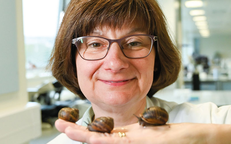
Principal Lecturer, School of Pharmacy and Biomolecular Sciences, University of Brighton
Investigation of the mode of action of aspernin, a novel antimicrobial protein identified in the mucus of the brown garden snail Cornu aspersum (Helix aspersa)
Working with her husband, Dr Alan Gunn of Liverpool John Moores University and other colleagues, Sarah’s research reveals that the defensive mucus produced by the brown garden snail, Cornu aspersum (also called Helix aspersa) has antimicrobial properties. Samples of the mucus were tested against a range of microorganisms, but a consistent, reproducible effect was only observed against Pseudomonas aeruginosa.
In disc diffusion plate antimicrobial assays, the mucus inhibited the growth of several type culture collection strains and clinical isolates of the bacterium. This is interesting because P. aeruginosa is a ubiquitous opportunistic pathogen. It infects a range of sites, including deep wounds and it is also an important cause of respiratory disease in patients with cystic fibrosis. Drug resistant strains are increasingly being reported, so a novel antimicrobial treatment would be valuable.
Using HPLC and mass spectroscopy the scientists were able to find partial sequences for three proteins with antibacterial activity. They have obtained the RNA transcriptome for the whole snail and used this to identify the full sequences for these three proteins. The one potentially of most interested in is a 37kDa protein, which they have named aspernin. The IBMS research grant will be used for the investigation of the structure, function and mechanism of antibacterial action of aspernin. Mucus is very difficult to work with, but now that they have the sequence, it will be possible to make the protein in a suitable vector. Having the protein on its own also allows quantitative assays, in particular the minimum inhibitory concentration of aspernin with respect to P. aeruginosa. The bacterium will also be grown in the presence of aspernin and the effect it has on the cell wall integrity, size and shape of the cells, will be examined using electron microscopy. Fluorescent-labelled amino acids will be used to see whether it affects protein synthesis.
A key experiment will be to test the effect of aspernin on mammalian cells using an in vitro cytotoxicity assay, to ensure that the protein could be suitable for use in human medicine. With results indicating the site and possible mechanism of action, it is hoped that aspernin could be used to develop a novel, clinically useful antibacterial treatment. It is envisaged as being used in combination therapies with existing antibiotics, in the form of a cream as a topical application for deep wound infections or an aerosol to treat respiratory infections.

Senior Lecturer at the University of Wolverhampton (BSc and MSc biomedical science degrees programmes).
Besides my teaching commitments, I run a laboratory developing improved treatments, disinfectants and diagnostic tests for Acanthamoeba keratitis (AK).
This is a potentially blinding infection of the cornea that is usually seen in contact lens wearers. The infection is caused by Acanthamoeba which is a protozoan organism that is widely spread in the environment and can be routinely isolated from sites around the home including tap water, garden soil and household dust.
Although a rare infection, AK is one of the most difficult infections to manage due to the absence of an effective monotherapy. Currently treatment times are protracted and range from six to 30 months, with up to 25% of patients requiring a corneal transplant and 5% requiring surgical removal of the eye.
Early diagnosis and instigation of treatment is required for a good prognosis and visual acuity post infection. However, no commercial diagnostic test exists for AK and currently diagnosis involves culturing corneal scrapes on non-nutrient agar plates seeded with an Escherichia coli food source. Unfortunately, this method takes 1-2 weeks and has poor sensitivity with a negative result in up to 50% of patients that actually have the infection.
Therefore, there is an urgent need for the development of a rapid and sensitive diagnostic test for AK that can be performed in diagnostic laboratories around the world. The project will be a collaboration between the University of Wolverhampton and Dr Steven Coles and Dr Gary Keane from the University of Worcester. On a previous project funded by the IBMS, the team produced the first monoclonal antibody for the detection of Acanthamoeba.
On this current project, the team is now going to incorporate the antibody into a lateral flow device (LFD) similar to a home pregnancy test, which will give a visual colorimetric result in two to five minutes. Initial evaluation of the LFD will then be performed using different strains of Acanthamoeba and fresh clinical isolates. If successful, this diagnostic test has the potential to dramatically speed up the diagnosis
of AK, resulting in an increased benefit to patients through reduced corneal damage and shorter treatment times.
Further information about IBMS research grants can be found on the IBMS website. The deadline for applications is 31 March in the year of application. To apply for an IBMS research grant download and complete the application form that can be found on the IBMS website.