Tracey de Haro, Specialist Scientific Lead for Electron Microscopy, University Hospitals of Leicester, looks at the revamped qualification.
The Diploma of Expert Practice (DEP) in Ultrastructural Pathology has been reviewed and updated by experts appointed by the IBMS. It has been re-introduced into the repertoire of professional qualifications of the IBMS to reflect a need within the scientific community to evidence and validate the knowledge and competence of scientists working in this specialised field. This qualification will support the career pathway of those working in electron microscopy (EM). This is particularly relevant as an increasing amount of diagnostic EM is being outsourced to units away from the originating trust.
Most diagnostic EM units are predominantly scientist-led with only the end product of the process being issued to a pathologist for consideration and inclusion into a diagnostic report. This is particularly pertinent to those units taking in referred work from outside their own hospital or trust. There is a high level of responsibility on the scientists working in this environment to get an accurate and considered answer in a timely fashion to the pathologist so that a final diagnosis can be made.
Even at the point of tissue dissection, decisions need to be made. Does the tissue require orientation, does it need cutting and how to do this without compromising the outcome? How can this tissue be processed to achieve the best ultrastructural detail?
Understanding
Once toluidine blue-stained semi-thin survey sections have been prepared they need to be assessed for the most appropriate area to be selected for ultrastructural examination. This requires knowledge of normal tissue morphology and a good understanding of pathological processes.
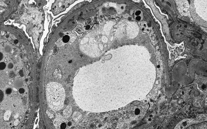
Understanding that a sclerosed glomerulus is not likely to give useful ultrastructural information or that if the clinical query is a mitochondrial disorder in a muscle biopsy, then you need to look for accumulations of dark blue material around the edge of a muscle fibre for example is vital. The sample size examined in the electron microscope is so small that it is very important to select the most informative area of interest to go through for ultrathin sectioning. This decision-making is done in some EM units by a pathologist but in many units, this is carried out by a scientist and carries with it a large degree of responsibility.
Examining
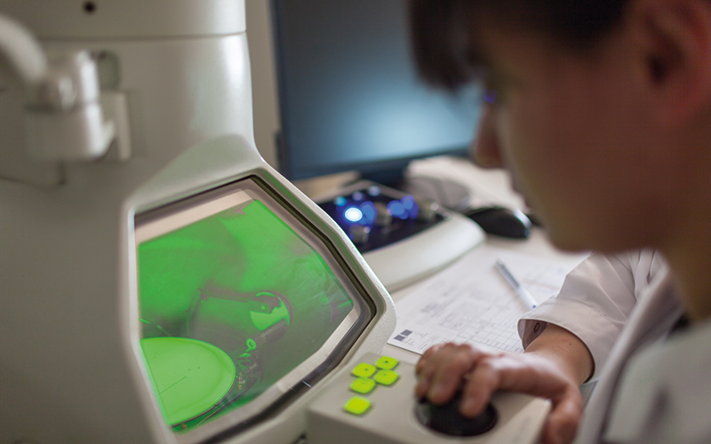
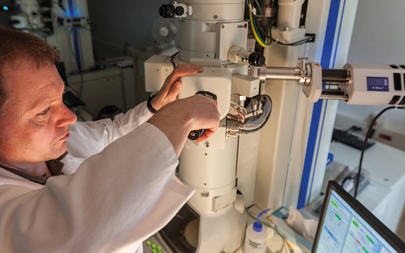
Examination of the ultrathin sections in the electron microscope is largely done by scientists, with images and ideally an ultrastructural report issued to the pathologist as the final product of this process. At this stage a high level of knowledge and understanding is required to ensure accurate and relevant information is relayed to the pathologist.
Examining a diagnostic sample on the electron microscope is more than just taking a set of appropriate representative images. It’s about considering the clinical information provided – what does that mean for the examination? For example, if the patient serology tests show an expression of a paraprotein then the renal tissue should be examined for a variety of possible disorders from non-organised immune complex deposits within the capillary loop basement membrane and tubular basement membranes, organised deposits within the glomerulus including amyloid and a variety of fibrillary disorders and finally proteinaceous casts with crystalloid inclusions within the tubules.
If the muscle biopsy has been taken for dermatomyositis, then tubulo-reticular inclusions need to be looked for within the endothelial cells of blood vessels. It’s also about the understanding that disease processes may not always be obvious or look the same in every case. If an IgA nephropathy is suspected for example, if only one glomerulus is examined and electron-dense deposit is not found then it doesn’t necessarily mean that the patient does not have an IgA nephropathy – just that a second or a third glomerulus needs examining as the disease process can be variable in presentation and has just not been found in that particular “slice” of the first glomerulus.
I could go on but the message here is that you must understand the tissue you are examining and be able to take into consideration all of the information you have at hand, such as clinical information, light microscopy results etc. alongside a good understanding of relevant pathology in order to make the right decisions about what to look for and image in your sample.
“The message here is that you must understand the tissue you are examining”
Going one stage further and adding an ultrastructural report to go alongside a set of relevant and appropriate images adds value to the process. A set of images gives a lot of information but it doesn’t give a full picture of what has been seen on the electron microscope. A set of images alone will not tell the pathologist that you have looked for tubulo-reticular inclusions and not found them or that the vacuolation seen was only found within a single muscle fibre. A good description can aid the pathologist to understand the images provided when they may well have not had prior involvement in the earlier EM process.
Apply for the Diploma

This qualification is intended for those scientists involved in imaging samples on the electron microscope and taking those decisions along the sample pathway. It also provides a good training plan for less-experienced staff to gain the necessary knowledge and experience of their colleagues and helps to ensure a resilience of service.
To apply for the DEP in Ultrastructural Pathology applicants must:
- be registered with the HCPC as a biomedical scientist
- have Membership (MIBMS) or Fellowship (FIBMS) of the IBMS
- have at least three years whole-time equivalent post-registration experience in histology
- have at least two years current practical experience in ultrastructural pathology.
The format of this DEP qualification follows that of the other DEP qualifications, such as the DEP in Histological Dissection, and involves a portfolio assessment and two written examinations that will cover the mandatory and optional modules.
The mandatory modules cover:
- Clinical governance
- General principles of electron microscopy preparation techniques
- Pathological process relevant to electron microscopy
- Electron microscope use
- Ultrastructural examination.
The optional modules cover the following tissue types:
- Skin
- Primary ciliary dyskinesia
- Muscle and nerve
- Renal biopsies.
Due to the workload pressures that many pathologists are under, most no longer have the time to view diagnostic samples on the electron microscope even if they have access to the transmission electron microscope (TEM) in their hospital.
They may be able to review particularly interesting or challenging cases, but many will rely on the scientist to provide an informed output. Pathologists who outsource their EM to a unit outside of their trust are entirely reliant on the knowledge and expertise of the scientists working in the referral EM unit.
Indeed, in some specialist areas, such as those examining cilia for primary cilia dyskinesia, pathologists play no part in this process, with scientists working directly with clinicians to achieve a diagnosis.
How do the pathologists and laboratories know that the scientists working in the EM unit carrying out their work are up to the job? How do they know that the scientists have the correct level of knowledge to be sure that the examinations and assessments carried out are done in a considered and informed way? Each EM unit will have competencies in place for the scientists, but these are local documents and will therefore vary between different units. There has been until now no common standard to competencies to prove the level that an individual is working at.
Evidence knowledge
The IBMS DEP in Ultrastructural Pathology will enable successful candidates to evidence the standard of knowledge and experience they have in this specific area in a way that will be recognised throughout the biomedical science arena. It is a way that the individual can validate their knowledge and experience – to prove they really do know what they are doing. It moves a conversation along from “yes, I can take images from a diagnostic sample” to “yes, I can take informed images from a diagnostic sample and give you an informed report on the ultrastructure that is relevant to diagnosis.”
It’s also a way that the wider department can validate to UKAS and other trusts sending work into an EM unit that their EM service is run by highly trained and qualified scientists – this is essential when scientists in most EM units work quite autonomously from others in their department.
It is already an expectation that scientists with responsibilities for the dissection of specific specimen types will have studied for and passed the
DEP in Histological Dissection and 45 candidates sat that exam in November 2022. Therefore, scientists working autonomously in EM should also be expected to be similarly qualified through this revised DEP in Ultrastructural Pathology.
For more information on the DEP in Ultrastructural Pathology, including the full Guidance to Candidates and Trainers, please visit the IBMS website or contact the examination team via [email protected]
Image credit | Shutterstock