Beginning her career as a junior hospital technician, Dr June Almeida completed it as a world-renowned virologist whose techniques revolutionised diagnostic electron microscopy. She was also the first person to see a human coronavirus. Here we look back over her life.
June Dalziel Hart was born on 5 October 1930, in a second-floor tenement flat at 10 Duntroon Street, Glasgow, her mother a shop assistant and her father a bus driver (her younger brother Harry died from diphtheria aged six years in 1940). Flourishing academically at Whitehill Senior Secondary School, Dennistoun, where she won a school prize in science, financial constraints meant that June could not go to university.
So, having passed her “higher” examinations, she left school in 1947 to become a junior histopathology technician at Glasgow Royal Infirmary, earning 25 shillings per week.
Early days
In 1952 the Harts moved to London, where June joined the pathology department of St Bartholomew’s Hospital as research assistant to Professor John WS Blacklock (1896–1973) and became an Associateof the Institute of Medical Laboratory Technology – the forerunner of the Institute of Biomedical Science.
In 1956, having married the Venezuelan artist Henry (Enrique) Rosalio Almeida (1913–1993) two years before, the Almeidas emigrated to Canada, where June was appointed as research assistant to Dr A F Howatson in the Division of Biological Research, Ontario Cancer Institute (OCI), Toronto, and her work with the electron microscope (EM) began.
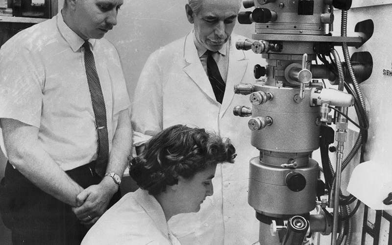
Electron microscopy was an emerging field of study in the 1950s, but Almeida’s expertise was apparent, and her reputation grew, with her first peer-reviewed journal article submitted for publication only one year after starting work in Toronto. In his entry in the Oxford Dictionary of National Biography, consultant virologist Professor Jangu Banatvala suggests that in Canada and the North American continent the lack of a university degree did not obstruct career progression, when compared with the UK.
“Electron microscopy was an emerging field in the 1950s, but Almeida’s expertise was apparent”
Please click here to read the full article.
Image credit |Getty| Alamy | Shutterstock | iStock