Specialist Biomedical Scientist Chloe Knowles looks at digital pathology and remote working during the pandemic.
On Wednesday 18 March 2020, the National Pathology Imaging Co-operative (NPIC) team came into the office for the last time that year. On hearing the Government’s advice that anyone who can work from home should do so, the team members set up in their own homes. The NPIC deployment team have planned and delivered the replacement of the existing digital pathology workflow in Leeds, and started new workflows with laboratories in the West Yorkshire region remotely, and it has not been without its challenges. This article describes how the deployment of digital pathology was done during the pandemic, and highlights some of the key factors that are crucial to a successful implementation, made even more so whilst working remotely. It will also cover the benefits of digital pathology, and how it has the potential to support remote working during the COVID-19 pandemic and the aftermath.
The first site
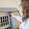
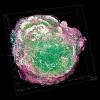
The histopathology laboratory at Leeds has been utilising digital pathology since 2018 and, as part of the NPIC, was the first site to go live with next-generation scanners and a new digital pathology image-viewing software. Leeds produces approximately 250,000 slides per year, equating to around one terabyte of image data generated every day. The NPIC is a unique collaboration between the NHS, industry and academia. The project began in 2019 and will deploy digital pathology solutions in over 40 hospitals across England, including sites within the West Yorkshire Association of Acute Trusts. This will create over 2.3 million images every year, and generate a staggering three petabytes of image data each year.