Could 3D imaging developments from the US change the way that diagnostic tests are carried out?
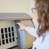
New technology that uses very simple optical hardware and a lens-free microscope, along with sophisticated algorithms, could make it easier and less expensive to diagnose chronic diseases. The new hologram system uses the technology to reconstruct images of tissue samples.
The system involves making biological samples transparent and then imaging them using a lens-free microscope. It could make much-needed diagnostic testing available and affordable for people in developing countries and remote areas that lack the expensive lab equipment currently used to perform tissue biopsies.
Gold standard
Tissue biopsy is widely considered the gold standard for detecting diseases, such as cancer and inflammatory conditions. But the test is relatively expensive and complex, and requires the use of sophisticated facilities — which can be a serious challenge in regions with limited resources, or remote areas.
In a standard biopsy, tissue is cut into thin slices – around one-tenth of the thickness of a human hair – and stained with dyes, so that medical professionals can use a microscope to detect abnormalities and diseased cells. One major limitation of that approach (beyond the time and cost involved) is that only a small number of tissue samples can be analysed at a time.
The new system was developed by a team led by Aydogan Ozcan and Rajan Kulkarni, from the University of California Los Angeles (UCLA). Aydogan says: “Although technological advances have allowed physicians to remotely access medical data to perform diagnoses, there is still an urgent need for a reliable, inexpensive means for disease imaging and identification – particularly in low-resource settings – for pathology, biomedical research and related applications.”
Cost saving
The researchers prepared tissue samples using a technique called Clarity, which makes tissue transparent, or “clears” it, using a chemical process that removes fat and leaves behind proteins and DNA. The method typically requires fluorescent dyes, which can be costly, and one drawback of the dyes is that the staining tends to degrade over time, making it harder for scientists to gather information from it.
But the UCLA researcher team used coloured, light-absorbing dyes, which it is claimed can be used with regular microscopy tools without any noticeable signal loss over time.
Instead of using the microscopes typically used for biopsy testing, the scientists developed a new device made of components that collectively cost just a few hundred dollars: a holographic lens-free microscope that’s capable of producing 3D pictures with one-tenth of the image data that conventional scanning optical microscopes need to perform the same function.
The UCLA method also allows the scientists to use tissue samples that are 0.2 millimetres thick – more than 20 times thicker than a typical sample. Again, they say this is a significant cost saving, as the sophisticated equipment required to produce thinner tissue slices is not required. This enables scientists to study a larger sample volume, which could help them to detect abnormalities earlier than they otherwise would.
The process
So how does the test work? Firstly, the cleared tissue is placed in a small container on a silicon chip that contains millions of photo detectors – the same type of chip that’s found in mobile phone cameras.
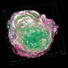
When light is shined on the tissue sample, low-resolution shadows from the tissue sample fall on the chip. Those shadows, created by the interference of light scattered by the sample, form holograms of the tissue sample.
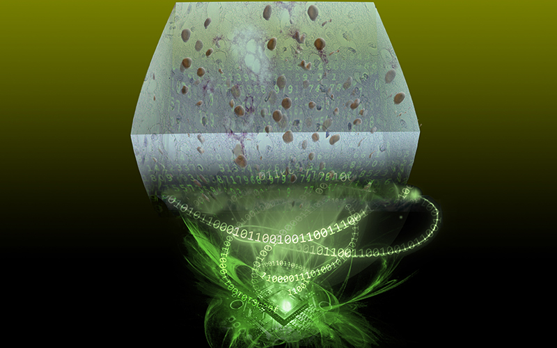
Next, the researchers enhance the resolution and enable 3-D imaging by shifting the sample relative to the image sensor and capturing the same holographic shadow, allowing them to digitally view different cross-sections, or digital slices, of the tissue sample.
Through computation and algorithms, the scientists converted a standard 10-megapixel imager, like those commonly used in mobile phones, into a few-hundred-megapixel microscope that can digitally image through different slices of a thick tissue sample.
The team has published a paper on this work, the abstract of which concludes: “Being low cost, simple, high-throughput, and data-efficient, we believe that this Clarity-enabled computational tissue imaging technique could find numerous applications in biomedical diagnosis and research in low-resource settings.”
Aydogan Ozcan is the UCLA Chancellor’s Professor of Electrical and Computer Engineering and Bioengineering and Associate Director of the California NanoSystems Institute. His work is supported by the Presidential Early Career Award for Scientists and Engineers, the Army Research Office, the National Science Foundation, the Office of Naval Research, the National Institutes of Health, the Howard Hughes Medical Institute, the Vodafone Americas Foundation and the Mary Kay Foundation
The scientists published their findings in Science Advances, a journal of the American Association for the Advancement of Science. For more information, visit bit.ly/BS_hologram