This series on tumour markers concludes with a review of pioneering work in the development, analysis and clinical application of an important serum tumour marker – prostate specific antigen.
There were 47,151 new cases in 2015 with 11,819 deaths due to PC. There is an increased risk of PC with age and over one third of cases are in the 65-74 age group
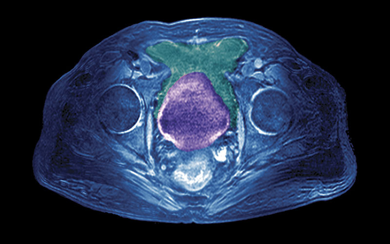
Prostate cancer (PC) is the most common cancer in males and constitutes around 26% of all cancer related deaths in the UK. There were 47,151 new cases in 2015 with 11,819 deaths due to PC. There is an increased risk of PC with age and over one third of cases are in the 65-74 age group, with a higher incidence in black males. Clinical features may be absent or include some of the following symptoms: urinary retention, frequency, nocturia, haematuria, impotence and weight loss meriting further clinical and laboratory investigation. The clinical process for diagnosis generally includes basal serum prostate-specific antigen (PSA), digital rectal examination (DRE) with palpation for any surface irregularities, especially a nodule, transrectal ultrasonography (TRUS) which may reveal benign prostatic hyperplasia, or suspicious hypoechoic areas which require TRUS guided needle biopsy and histological interpretation for a Gleason score. With a strongly positive diagnosis, an early isotopic bone scan may be performed. Around 20% of patients with PC have metastases at diagnosis with bone, liver or lung secondaries most common, with a five-year survival rate of around 29%, this increases to almost 100% if the detected tumour is local without metastases.
An early prostate biomarker
In 1935, Kutscher and Wolberghs at Heidelberg University reported that prostatic tissue and seminal fluid contained a high concentration of a phosphatase with optimal activity between pH 4.5-6. Alexander and Ethel Gutman adapted the King Armstrong colorimetric method for alkaline phosphatase with an acid buffer and found that with a cut off of 4 units % in a small study serum acid phosphatase was significantly raised in 11/15 patients with metastatic PC. The findings were confirmed in later larger studies by Sullivan (1942) and Herbert (1946). It is quite remarkable that this procedure with small variations was the method of choice for the clinical chemical laboratory investigation of PC for over four decades.
Prostatic acid phosphatase
An acid phosphatase fraction (PAP) from prostate tissue was reported by a New York State University research team, with lead author Sidney Schulman in 1964, using radial immunodiffusion. This led to quantitative immunoassays notably by Foti (1977) and Griffiths (1980) using solid phase radioimmunoassay, which showed improved diagnostic accuracy compared to the colorimetric enzyme assays for serum acid phosphatase (SAP). Serum PAP has been assessed and generally regarded as a highly specific but low sensitivity tumour marker, especially in the early stages of PC.
Prostate-specific antigen
The discovery of PSA in human prostate tissue extracts in 1970 is attributed to the American immunologist Richard Ablin, who used gel diffusion precipitation to identify prostatic antigens. It would be a further eight years before PSA was isolated and characterised in human seminal plasma as a potential forensic marker for rape crimes. Progress was achieved a year later at Roswell Park Comprehensive Cancer Center, New York with a research team led by T Ming Chu. PSA was identified and purified in 1979 and shown to be immunologically distinct from PAP. A monospecific antiserum to PSA was produced using tumour extracts as reactive immunogens. After studies using rocket immunoelectrophoresis, a sensitive, sandwich type enzyme immunoassay was developed to measure serum PSA, which was clinically assessed and found to be useful in predictive prognosis, monitoring treatment and the detection of PC recurrence.
Methods to measure
The Roswell Park cancer research team’s patent allowed biotechnology companies to produce PSA test kits from 1986 with FDA approval in the US, leading to their widespread use in the clinical setting. Kits, such as Tandem E PSA employed a solid phase-two site immunoenzymatic sandwich assay with two murine monoclonal antibodies and a chemiluminescent detection of substrate product. Another popular early kit assay was Pros-Check, used conventional radioimmunoassay with a rabbit polyclonal antibody, but results were typically almost twice that of the Tandem E assay. Concerns regarding calibration and reference ranges led to comparison studies, such as Kort and colleagues in 2006 for six automated immunoassay methods were found to show good comparison and close agreement with the WHO 96/ 670 international preparation. The generally accepted reference range for serum total PSA is 0-4ng/ml, but this is now more appropriately age related and of note the upper limit is raised to 6.5ng/ml in men 70-79 years of age.
Serum PSA
PSA is now identified as a single chain 33Kda glycoprotein of 237 amino acid residues with 4 carbohydrate side chains. It is an androgen, regulated, serine protease enzyme which is produced by normal prostate luminal epithelial cells. PSA is related to kallikrein serine proteases and can also be identified as hK-3. The main function of PSA is to liquefy the seminal coagulum to promote the release and motility of spermatozoa, some PSA escapes from the prostate and can be found in serum. Various molecular forms exist but PSA is released into blood with around 70% as a stable complex with alpha1 antichymotrypsin (CPSA) and free non-complexed PSA (FPSA) typically constitutes 5-40% of total serum PSA measured. The development of antibodies specific for CPSA and FPSA in 1991 led to immunoassays for each fraction and the use of percentage and ratios.
Clinical use of serum PSA
Serum PSA has several significant limitations as an ideal tumour marker. Levels are also raised in benign prostatic hypertrophy (BPH), prostate inflammation and infection and may be increased in other cancers, notably breast and kidney, and so it is not specific to the prostate. Serum PSA does not always correlate well with the aggressive nature of the tumour, raised results in patients with low grade tumours, which remain localised means treatment may be inappropriate, actually cause harm and affect quality of life. In addition, results are affected by some types of medication, for instance LHRH agonists/antagonists may reduce serum PSA by 50%. Other factors include age, race and weight and calibration and performance characteristics of the chosen method of analysis.
Screening
This has been the subject of numerous studies and trials. The 1986 FDA notice recommended serum PSA for monitoring only but with reliable assays available, its use in screening grew rapidly encouraged by observations that a fall in PSA following hormone therapy correlated with response to treatment and that increased results after treatment predicted recurrence. Many large scale clinical studies would take place, however, the chosen parameters often varied, notably in the upper decision limit of the reference range, use of DRE and core biopsies in follow up. It became recognised that patients diagnosed by screening had clinically organ confined disease and around 25% with PSA >4ng/ml were found to have PC on biopsy. Modifications were introduced, including age adjusted ranges, PSA velocity-measures rate of increase, PSA density – prostate size, and the use of free to total PSA %, which was found to be inversely related to the risk of PC.
The contribution of PSA screening in decreasing disease-specific mortality was in doubt due to the conflicting results from two major prospective prostate screening trials, which reported their findings in 2009. The European randomised study of screening for prostate cancer (ERSPC) presented data that showed a 20% decrease in the prostate specific mortality rate, while the Prostate, Lung, Colorectal, and Ovarian Cancer screening trial demonstrated no significant decrease in prostate specific mortality. Also of great concern was that each study showed that PSA screening led to overdiagnosis and overtreatment with unnecessary biopsies, treatment side effects and stress to the patient. PSA may detect small, low grade and localised tumours which may be clinically insignificant and not require treatment.
Prognosis and recurrence
Research described earlier showed that PSA may be useful in treatment monitoring, risk stratification, predictive prognosis and the detection of tumour recurrence. In 2008, the National Academy of Clinical Biochemistry (NACB) published recommended practice guidelines that PSA is useful for detecting disease recurrence and monitoring therapy and that free PSA is valid to distinguish malignant from BPH when total PSA is <10ng/ml. Numerous alternative prostate tumour markers, such as kallikrein 2 and myeloid protein-14, are still under evaluation. The clinical decision cut off point of <4ng/ml is endorsed by many of the expert panel groups. Irrespective of which type of treatment has been used from active surveillance, surgery, radiotherapy, chemotherapy or anti androgen therapy, regular measurements of serum PSA remain the yardstick for success or failure of treatment.
Active surveillance may involve serial testing of serum PSA every six months, with DRE and an annual biopsy, which, based on results, may lead to treatment. It has also been claimed that a single PSA in early middle age can predict risk of advanced prostate cancer decades in advance, which can help to plan future screening. Expected PSA results following successful TURP (transurethral resection of prostate) at one month are <0.2ng/ml, which requires using an ultrasensitive PSA assay. However, a rise within next 12 months, or a doubling time of six months, indicates progressive disease. The preoperative PSA and the interval between surgery and redetection of PSA by conventional assay can be used to predict disease free survival and pattern of recurrence. Following radiotherapy the decline in PSA is less and lowest level may require at least 12 months, but a rise >2ng/ml over lowest result or three consecutive rises at three and six month intervals tends to indicate radiation failure.
Concluding comments
Despite its controversial role in screening, serum PSA has been the mainstay biochemical marker in prostate cancer for over 30 years. Advances in treatment, such as higher dose radiotherapy, and imaging techniques with increased government funding may improve the outlook for prostate cancer patients.
Stephen Clarke is a retired IBMS Fellow.
Picture credit | Science Photo Library
References
1. Cancer Research UK data 2015
2. Hamilton W, Sharp DJ, Peters TJ et al. Clinical features of prostate cancer before diagnosis: a population based, case control study. Br J Gen Pract 2006; 56(531): 756-762.
3. Gutman AB, Gutman EB. An ‘acid’ phosphatase occurring in the serum of patients with metastasizing carcinoma of prostate gland. J Clin Invest 1938; 17(4): 473-478.
4. Schulman S, Mamrod L, Gonder MJ et al. The detection of prostatic acid phosphatase by antibody reaction in gel diffusion. J Immunol 1964; 93: 474-480.
5. Griffiths JC. Prostate specific acid phosphatase: revaluation of radioimmunoassay in diagnosing prostatic disease. Clin Chem 1979; 26(3): 433-436.
6. Kurijama M. Prostate specific antigen as a tumor marker in prostate cancer. Int J Urol 1994; 1: 99-113
7. Ablin RJ, Soanes WA, Bronson P et al. Precipitating antigens of the normal human prostate. J Reprod Fert 1970; 22: 573-574.
8. Sensabaugh GF, Crim D. Isolation and characterisation of a semen specific protein from human seminal plasma: a potential new marker for semen identification. J Forensic Sc 1978; 23: 106-115.
9. Diamandis E, Bast R, Gold P, Ming Chu T, Magnani JL Reflection on the discovery of carcinoembryonic antigen, Prostate specific antigen, and cancer antigens CA125 & CA 19-9 Clin Chem 2013; 59(1) : 22-31.
10. Kurijama M, Wang MJ, Papsidero LD et al. Quantitation of prostate-specific antigen in serum by a sensitive enzyme immunoassay. Cancer Res 1980; 40: 4658-4662
11. Kort SAR, Martens F, Vanpoucke H et al. Comparison of six automated assays for total and free PSA with special reference to their reactivity toward the 96/670 reference preparation. Clin Chem 2006; 52(8): 1568-1574.
12. Duffy MJ, McGing P. Guidelines for the use of tumour markers. ACB (Ireland) 4th edition 2010, p17-21
13. Bryant RJ, Lilya HG. Emerging PSA-based tests to improve screening. Urol Clin N Am 2014; 41(2): 267-276
14. Pavlou MP, Diamandis E, Blasutig IM. The long journey of cancer biomarkers from the bench to the clinic. Clin Chem 2013; 59(1): 147-157
15. Hernandez J, Thompson I. Prostate specific antigen: a review of the validation of the most commonly used cancer biomarker. Cancer 2004; 101(5): 894-904
16. Partin AW, Brawer MK, Bartsch G et al. Complexed prostate specific antigen improves prostate cancer detection: results of a prospective multicentre clinical trial. J Urol 2003; 170(5): 1787-1791
17. Jung K, Elgeti U, Lein M et al. Ratio of free to complexed prostate specific antigen to total PSA: which ratio improves differentiation between benign prostatic hyperplasia. Clin Chem 2000; 46(1): 55-62
18. Schroder FH, Hugosson J, Roobool MJ et al Screening and prostate cancer mortality in a randomised European trial. New Engl J Med 2009; 360: 1320-1328
19. Andriole GL, Crawford ED, Grubb RL III et al Mortality results from a randomised prostate cancer screening trial. New Engl J Med 2009; 360: 1310-1319
20. Sturgeon CM, Duffy MJ, Stenman UH et al. National Academy of Clinical Biochemistry practice guidelines for the use of tumour markers in testicular, prostate, colorectal, breast and ovarian cancers. Clin Chem 2008; 54(12): e11-e79
21. Botchorishvili G, Matikainen MP, Lilya H. Early prostate specific antigen changes and the diagnosis and prognosis in prostate cancer. Curr Opin Urol 2009; 19(3): 221-226
22. Duffy MJ Tumour markers in clinical practice: A review focusing on common solid Tumours. Med Princ Pract 2013; 22:4-11
23. Kurijama M. Prostate specific antigen as a tumour marker in prostate cancer. Int J Urol 1994; 1: 99-113
24. NICE CG175 clinical guidance prostate cancer: diagnosis and management January 2014.
25. Ellis WJ, Vessella RL, Noteboom JL. Early detection of recurrent prostate cancer with an ultrasensitive chemiluminescent prostate specific assay. Urol 1997; 50(4): 573-579
26. Pound CR, Partin AW, Eisenberger MA. Natural history of progression after PSA elevation following radical prostatectomy. JAMA 1999; 281(17): 1591-1597