Nicki Lawrence, Principal Biomedical Scientist and Advanced Practitioner, introduces “Bite-Size Morphology” and presents a number of cases.
Morphology, in some laboratories, is thought to be a dying art with newer investigations such as immunophenotyping and cytogenetics replacing expert review of the peripheral blood film. As a consequence, there may be deskilling of biomedical scientists as the focus is shifted to these newer techniques when making a definitive diagnosis. Coupled with old-school morphologists retiring, this has led to a lack of experienced morphologists within some departments. Morphology, though, is still needed to make a working diagnosis; it’s quick, relatively cheap to make a blood film and within an hour of the sample arriving in the laboratory, you can make a preliminary diagnosis on a wide range of disorders, including life-threatening conditions such as acute promyelocytic leukaemia or microangiopathic haemolytic anaemia.
As an Advanced Practitioner in Morphology and our departmental Haematology Training Officer it is unsurprising that I am hugely passionate about morphology and education. Each week I present at our Clinical Haematology Morphology MDT Meeting and I wanted to provide something similar for biomedical scientists within our department, exposing them to a wide range of cases, both routine and more obscure, so that they can develop their morphology skills further. With the introduction of pathology networks across England I also wanted to extend this to our network laboratories in Leighton and Macclesfield.
I started presenting “Bite-Size Morphology”in July 2022 and decided on a 30-minute format during the working day, which was available to all staff via Teams. By using Teams I’m able to record each session and save them on our network drive so that anyone who is not able to attend can watch again later. Each session is themed, with three or four cases reviewed at a time.
We’ve covered a variety of morphology, including lymphocytes with cytoplasmic projections or prominent nucleoli, eosinophilia, polycythaemia vera, leucoerythroblastic blood films and the underlying cause, essential thrombocythaemia, and parasites including Loa Loa, Plasmodium falciparum and Chagas disease to name a few. In each session
I provide the history of the patient but we also discuss the pathophysiology behind each diagnosis with a question and answer session to end. All staff can email me and suggest future topics too or ask questions without an audience. At the end of the year I provide a CPD certificate stating which sessions were attended or watched. Particularly interesting cases are discussed in-depth in my monthly lunchtime seminars.
I’m determined to keep our workforce skilled in morphology and as we continue through 2023 I’ll be sharing more of our Bite-Size Morphology cases via my twitter account @Promyelocyte13 so that other biomedical scientists can also see a host of interesting cases. For now, presented here are four morphology case studies.
Case Study 1
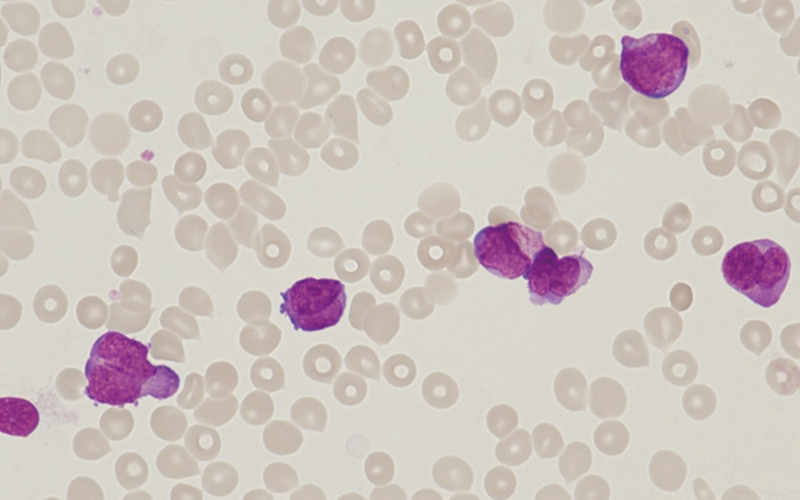
A 71-year-old male presented with shortness of breath.
Numerous abnormal promyelocytes, some with Auer rods, can be seen on the image (x600 magnification). The blood film was in keeping with a diagnosis of acute promyelocytic leukaemia (APL). Urgent FISH for PML::RARA was sent but no evidence of PML::RARA or a RARA gene rearrangement found. No evidence of an insertion/duplication within NPM1 gene found but 21bp and 45bp internal tandem duplications within FLT3 found (FLT3-ITD).
As the morphology was consistent with APL, further investigations were undertaken showing a cytogenetically cryptic PML::RARA gene rearrangement and the patient classified as having APL with PML::RARA fusion.
WBC 54.3 x 10⁹/L
Hb 111 g/L
Platelets 34 x 109/L
Case Study 2
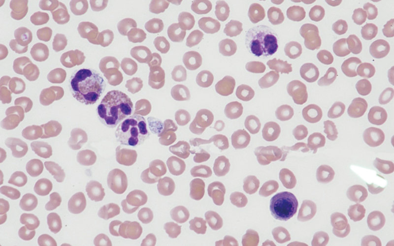
A 63-year-old female with a history of autoimmune haemolytic anaemia controlled with steroids presented with mild eosinophilia, which rapidly increased. The patient was admitted to RSUH from local DGH and transferred to ITU for multiple pulmonary emboli, pleural effusions, Stevens-Johnson syndrome (SJS) and suspected haematological malignancy. Patient was later confirmed as Stevens-Johnson syndrome, ovarian cancer and a clinically significant variant associated
with myeloid neoplasia was found in DDX41. Image (x600 magnification) shows multiple eosinophils with some degranulation and a single nucleated red blood cell.
WBC 50.8 x 10⁹/L
Hb 84 g/L
Platelets 17 x 109/L
Neutrophils 10.67 x 10⁹/L
Eosinophils 34.53 x 10⁹/L
Case Study 3
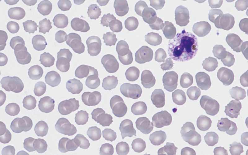
A 90-year-old female presented from her GP with severe headaches controlled by paracetamol.
The image (x600 magnification) shows thrombocytosis with platelet anisocytosis (variation in platelet size) and large and giant platelets can be seen. Based on the FBC results, normal inflammatory markers, normal iron studies, and blood film morphology, it was highly suggestive of a myeloproliferative neoplasm and I suggested referral to the Clinical Haematology department for further investigation. Initial screening for JAK2 V617F mutation was negative and the extended MPN panel was instigated with MPL and CALR mutations also tested. The patient was positive for MPL mutation.
WBC 8.4 x 10⁹/L
Hb 104 g/L
Platelets 1089 x 109/L
CRP <4 mg/L
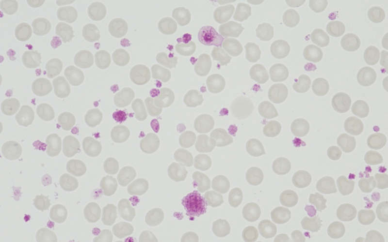
A 64-year-old male returned from Ghana one day previously presenting with headache, aches, fever and chills, now with shortness of breath. He did not take any malaria prophylaxis. Past medical history of splenectomy for road traffic collision. The top image (x600 magnification) shows numerous red cells containing Plasmodium falciparum trophozoites with a parasitaemia of 8.8% confirmed by London School of Hygiene and Tropical Medicine. The single neutrophil present contains malaria pigment. There are also some echinocytes present (red blood cells with evenly distributed frilly projections) in keeping with the patient’s acute renal failure.
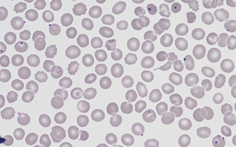
The patient was treated with artesunate and the usual mode of parasitised red cell clearance – splenic pitting – was not possible due to his previous splenectomy. The parasites, therefore, remained within the peripheral blood but the morphology changed. The bottom image (x600 magnification) shows shrunken trophozoite cytoplasm with only the chromatin dot visible in the majority of the trophozoites.
Nicki Lawrence is Principal Biomedical Scientist Advanced Practitioner in Morphology and Haematology Training Officer at University Hospitals of North Midlands NHS Trust.
Image credit | iStock