Scanning technology has identified hidden abnormalities in the lungs of long COVID patients.
Early on in the pandemic, Professor Fergus Gleeson, Consultant Radiologist at Oxford University Hospitals NHS Foundation Trust, had the idea to use a scanning method to understand changes in lung physiology due to COVID-19. The non-invasive method of hyperpolarised xenon MRI scans has been used for some years to develop understanding of lung physiology in health and disease, and was pioneered by Professor Jim Wild and the Pulmonary, Lung and Respiratory Imaging Sheffield (POLARIS) research group at the University of Sheffield.
Both Gleeson and Wild are among the research team in the EXPLAIN project – one of 19 studies into long COVID to receive £1.8 million government funding through the National Institute for Health Research, which uses hyperpolarised xenon MRI scans to look at ongoing lung damage in non-hospitalised COVID-19 patients. The project aims to improve understanding of long COVID, from diagnosis and treatment through to rehabilitation and recovery.
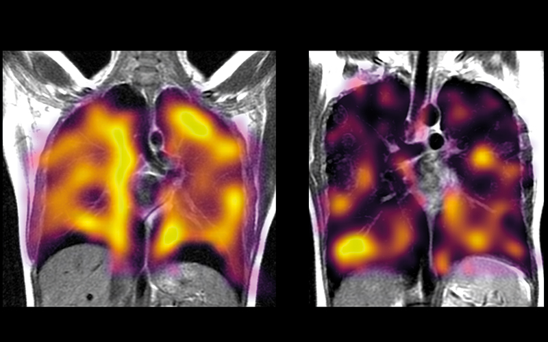
Evidence has emerged in the latest pilot study from EXPLAIN, of lung abnormality in long COVID patients undetected by other scans. Participants lie in an MRI scanner and inhale one litre of xenon, which has been hyperpolarised so it can be seen using MRI. It takes only a few minutes and, because radiation exposure is not needed, can be repeated over time to see changes to the lungs. As xenon follows the pathways of oxygen when it is taken up by the lungs and can show where the abnormality lies between the airways, gas exchange membranes and capillaries in the lungs, radiologists can observe how the gas moves from the lungs into the bloodstream.
A 2021 EXPLAIN study that scanned people who had been hospitalised due to COVID-19 established that they had persistent lung abnormalities several months after being discharged. Now a new EXPLAIN pilot study has investigated possible lung damage in COVID-19 patients who have not been hospitalised but have experienced breathlessness months after a diagnosis.
Hidden impairments
“Initially, we thought that there wouldn’t be any significant changes in lung gas transport, in comparison to healthy non-infected individuals, but how wrong we were,” says Dr James Grist, post-doctoral research scientist at the University of Oxford, and one of the researchers in the EXPLAIN study. “We were very surprised to see xenon gas transfer abnormalities in patients whose lungs otherwise looked healthy on conventional imaging.”
Transfer of gas from the lungs to the bloodstream was “significantly impaired” in those study participants who have been diagnosed with long COVID, seen in long COVID clinics and who have had normal computerised tomography scans. “The extent of the changes in gas transfer between healthy participants and those who have had a COVID-19 infection was really surprising,” Grist says.
“When we got our images from the first patient, I thought I had really messed up the data acquisition. However, we had a volunteer lined up afterwards and their images were fine. The differences really are that stark.”
Other infections
The pilot study had 36 participants – all recruited from the Oxford Post-COVID Assessment clinic – including people diagnosed with long COVID in clinics, those who had been hospitalised with COVID more than three months ago and those who had not been experiencing long COVID. All had normal CT scans.
The full EXPLAIN study will recruit about 400 participants, some of whom will have had breathlessness, some who have had COVID-19 but no symptoms, some with no breathlessness but other long COVID symptoms – such as “brain fog”, and some who have never had long COVID. “The next steps are to scan a lot more patients,” Grist says.
The research team is running a multi-centre study with the Universities of Sheffield, Cardiff and Manchester with multiple cohorts of patients to really “dig down into the details of what we are seeing here in our initial study”, Grist says. “We are currently assessing the correlation between our imaging results and the persistent breathlessness that some patients experience. ”
2011–2014 BSc Physics with medical physics, University College London
2010–2014 Dialysis auxiliary nurse specialist, University Hospital Birmingham
2013–2014 Research assistant in acousto-optics, University College London
2014–2018 PhD, multi-nuclear MRI, including carbon and sodium, University of Cambridge
2018–2020 Research fellow, University of Birmingham
2020–present Research Fellow, University of Oxford and Nicholas Kurti Junior Research Fellow, University of Oxford.
Future research
The next important questions to answer include how many patients with long COVID will have abnormal scans, as well as the significance of the abnormalities detected, their causes and longer-term consequences. Understanding the mechanisms that resulted in these symptoms should lead to the development of more effective treatments for long COVID, the researchers say.
“We really need to perform these studies to see how the lung responds to infection after other respiratory illnesses, such as influenza, to understand whether these changes are unique to COVID-19 or not,” Grist continues. “We’ve love to expand this study to patients who have had other seasonal respiratory infections too.”
Grist’s career has included being in the team to conduct the first human studies using the imaging technique hyperpolarised carbon-13 MRI to better understand multiple sclerosis, using machine learning to predict survival in childhood brain tumours and hyperpolarised xenon MRI in long COVID. “It really has been a pleasure to work with fantastic colleagues, both staff and students, and I am really proud of our accomplishments. Science really is done best in a team,” he says.
“I cannot put my current career success down to anything except that I have been able to work with some really talented scientists in the right place at the right time.”
The imaging techniques are ground breaking, and, he concludes, “have the potential to both push forward our knowledge of physiology, advance radiology practice, and really make a difference in the lives of patients across a wide spectrum of the healthcare system”.
Image Credit | NIHR Oxford Biomedical Research Centre BRC | Getty