Kinjal Patel, Advanced Practitioner in Histological Dissection, gives a guided tour of her lab at Charing Cross Hospital.
I am based in the cellular pathology department at Charing Cross Hospital, which is managed by North West London Pathology. This is an NHS pathology partnership between Imperial College Healthcare NHS Trust, Chelsea and Westminster NHS Foundation Trust and Hillingdon Hospitals NHS Foundation Trust (which comprises nine hospitals in total) delivering safe, high-quality and reliable care to local communities and residents.
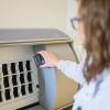
This department incorporates cytopathology, histopathology, electron microscopy, and specialised integrated haematological malignancies diagnostic services. We are a specialised diagnostic centre for trophoblastic diseases and also provide comprehensive diagnostic services including immunohistochemistry, immunofluorescence, MOHS service and frozen sections and medical renal diagnostic services. In March 2020, the hub laboratory for histopathology at Charing Cross was completed, which included the harmonisation of services and implementation of new state-of-the-art equipment and IT systems.
We are a high-performing team and pride ourselves on the quality of service that we offer. Our workforce consists of biomedical scientists, clinical scientists, non-qualified staff and pathologists. Our laboratory is IBMS training approved, offering training and development in the form of pre- and post-registration opportunities, including dissection and reporting, among many others. Staff are rotated between sections, promoting and supporting development and growth of professional skills to sustain their interest in the field.
Our quality management system incorporates comprehensive internal quality assurance and control procedures. We participate in relevant UKNEQAS schemes, and other external quality assurance schemes. To provide continuous improved service we perform numerous internal audits that allow us to reflect and review our procedures and to identify gaps in our processes. During the current global pandemic, and straight after our services moved to the hub laboratory, we achieved outstanding accreditation by UKAS to the ISO 15189 standard without any findings.
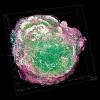
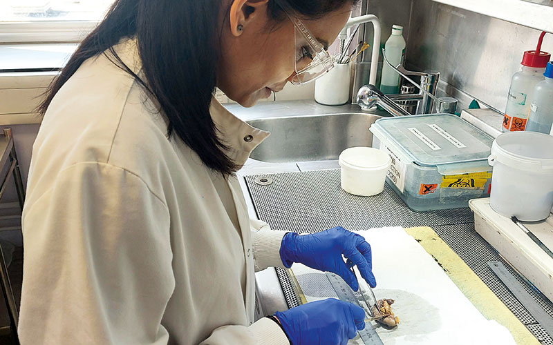
My main duties focus on our specimen reception area, where we process a variety of specimens, from biopsy to complex resections. As one of the largest NHS cellular pathology laboratories in the UK we have a huge specimen reception with nine dissecting benches, six specimen dissectors and two advanced practitioners working towards reporting gastrointestinal and gynaecological histopathology. I am currently undertaking my dissection portfolio, which is something I would not be able to do without the support network within the department.
Although the last year has been challenging, I have witnessed the determination of our agile team to provide a high-quality diagnostic service able to adapt and implement new ways of working.
Working in a complex department with a huge workload, sometimes it is difficult to stop and reflect. However, I witness my colleagues rising to new challenges every day while still upholding stringent quality and health and safety standards. In addition our management team implemented procedures to ensure that our teams could work safely in a nourishing environment keeping patient care at the centre of everything.
Our hope for the future is to continue to develop and improve our service by recruiting more staff, investing in innovative new technology and promoting the vital work that all pathology laboratories provide in order to deliver the best patient care we can to the communities in our area.