Former Chief Biomedical Scientist Ivor J Mitchelmore outlines a trip to India, where he and colleagues worked to set up a microbiology culture service.
Earlier this year, along with two ex-microbiology colleagues, Hilary Rogers and Ruth Parry, I visited a small mission hospital in the village of Sarenga, West Bengal. It is called Khristiya Seva Niketan (KSN) hospital and serves a large rural population, mainly of tribal people.
Maternity and children make up the majority of inpatients, plus there are busy outpatient clinics. While we were at KSN, they held an eye camp, at which over 40 cataract operations were performed. Building work has started for a blood bank, which will enable KSN to perform more complex surgery. The hospital already has a small pathology laboratory offering a basic range of haematology and biochemistry tests, but no microbiology culture service. This means that patients with suspected infections are treated blind, with nothing to say whether the treatment is appropriate other than observing their condition.
Better prepared
On a visit to the hospital in 2018, the Medical Superintendent and other doctors at the hospital said that they would value the setting up of a microbiology service. Having had a disappointing experience last year, when I spent the first three months at KSN trying to help them set up microbiology testing, I was determined this time.
The three of us all had our maximum luggage allowance of 40kg each, and over half of this was due to donations of equipment and consumables from companies and local laboratories.
I had earlier shipped out a benchtop autoclave, which proved invaluable. Yes, you can buy an autoclave in India, but will it work? After trying three last year, all of which failed, I decided to take one that I knew would do the job.
Hilary had recently retired after 37 years service at the Luton and Dunstable Hospital, and is an expert in media production (along with bacteriology, mycology and parasitology). Within a few days, we had a fridge full of culture media. This was almost entirely produced by the local staff under Hilary’s supervision. I was thankful that we did not have to resort to bleeding the staff to make the blood agar plates (as happens at another hospital I have visited in India), but instead we managed to obtain an expired pack of blood from a blood bank.
Obtaining samples
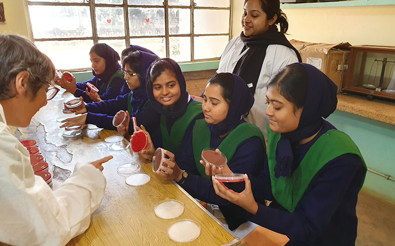
We had taken a few blood agar and Mueller-Hinton agar plates with us, and immediately set up nose swabs from the lab staff, in the hope of obtaining a Staphylococcus aureus control. We drew a blank there, but then had the idea of swabbing all the 25 first-year nursing students from the nurse training school attached to KSN. This turned into a lab-based training session for the students, who have a large microbiology unit in their first year. It was great fun as they took each other’s nose swabs and then handed them to Ruth for plating out.
Sure enough, when the students came back the next day, several of their nose swabs had grown S. aureus, as confirmed by the staph latex kit and rabbit plasma we had brought with us. One girl was horrified to discover she had S. aureus in her nose, and we explained to her that it was normal for some people.
For training purposes we inoculated a rectal swab onto a range of plates, which served to demonstrate Proteus swarming, and provided a Pseudomonas and a lactose-fermenting coliform as control organisms. We also isolated our first anaerobic bacteria, using an anaerobic jar.
To create a CO2-enriched atmosphere, we used a candle in a sealed biscuit tin. From a catheter specimen of urine we were able to isolate Group B beta-haemolytic streptococci from the mixed growth. We now had positive and negative control organisms for slide and tube coagulase, staph latex, oxidase and catalase tests plus the Group B streptococci to control the strep latex procedure – very important to enable local staff to practise their newly learnt skills. With the control organisms we had gathered, Ruth organised training the staff in making and reading Gram-stained films. With limited resources, this will be a key step in identifying their isolates.
The most satisfying result was when the obstetrics and gynaecology doctor sent a urine sample from a patient visiting the weekly antenatal clinic. It had a high white cell count and the following day we had grown what looked like a fully sensitive Escherichia coli. The same result would have taken five days if we had sent it to an outside laboratory. This was processed by local staff and the patient was started on nitrofurantoin, but later admitted and changed to IV gentamicin. She made a complete recovery.
We have since grown urine isolates that have been resistant to five out of six antibiotics tested, so it shows the importance of checking the susceptibility patterns of these organisms.
Tremendous progress
As well as the practical work I gave regular half-hour presentations to staff, explaining the theory behind what they had been learning at the bench. I don’t speak much Bengali, so there was a language barrier, but the staff all speak some English and the younger ones are very good at explaining things to the others, where necessary.
Ruth and Hilary returned to the UK after two weeks, but I could not have achieved the tremendous progress we have made without them, or without the generous contributions from the trade and local UK laboratories (see box). I had to keep reminding myself that in seven weeks we were expecting the local staff to pick up skills that took me a year to begin to get to grips with. The lab staff at KSN have done incredibly well, but still need a lot of support.
Time will tell how effectively this can be delivered via WhatsApp. I am already thinking about a November visit to give some booster sessions, and take them to the next level. At least now I have the confidence to know that change is possible.
Contributions
Ivor J Mitchelmore is a former Chief Biomedical Scientist in the department of medical microbiology at Luton and Dunstable Hospital.
He would like to thank the following companies and laboratories for their contributions to the project: BioConnections, Don Whitley Scientific, Mast Group, Medical Wire, Pro-Lab Diagnostics, Department of Medical Microbiology at Luton and Dunstable Hospital and the Department of Microbiology at Watford General Hospital.