News

AddToAny

Google+ Facebook Twitter Twitter

Imaging with LEGO

Researchers have come up with an inexpensive, automated way to image biological samples – using the children’s toy, LEGO.

p8-11-news-lego-blocks-istock-459012875.jpg

In a new paper scientists at the Crick, UCL and Aix University in Marseille describe a novel device, built with LEGO parts, that can be adapted to most microscopes.

Project lead Ricardo Henriques said: “Sometimes the best solutions to complex biological questions are remarkably low-tech. By developing hardware that uses inexpensive LEGO components as its building blocks, we’ve built an accessible tool that delivers customisable cutting-edge microscopy at a fraction of the cost.”

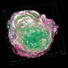
The device, called NanoJ-Fluidics (and nicknamed Pumpy McPumpface), enables researchers to observe cells in a highly customisable format. For the very first time, researchers can observe samples of cells at well-defined moments in time, for example when they are dividing or become sick.

The LEGO hardware and accompanying software are fully open-source, enabling other research groups to make their own devices. NanoJ-Fluidics has already been successfully adopted by over 10 labs across the world.  

Image credit | iStock

Related Articles

illustration_CREDIT_istock-484714523

Advanced and consultant practice roles

Sarah May and Chris Ward from the IBMS look at advanced and consultant roles and discuss new developments and qualifications.

Robotic hand with handgun aiming in wrong direction. Fatal AI error - Image credit - Shutterstock-2309476357

Tissue contamination distracts AI

In a new study, scientists trained three AI models to scan microscope slides of placenta tissue to detect blood vessel damage, estimate gestational age and classify macroscopic lesions.

Digital pathology cleared for use-history digital pathology-CREDIT-university of warwick

Digital pathology cleared for use

New research has led to the UK government approving the use of digital pathology to help speed up analysis of cancer screening samples.

Breast cancer 3D tomography-CREDIT-Science-Photo-Library-c0303374

Two decades of hHER2 testing

This year marks the 20th anniversary of Her2 breast cancer testing at the Royal Cornwall Hospital. Senior Biomedical Scientist Mary Jones looks back over the past two decades.

Top