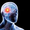
Researchers from Japan may have devised a way to perform the initial biopsy, lab tests, and subsequent tumour removal during one surgical procedure.
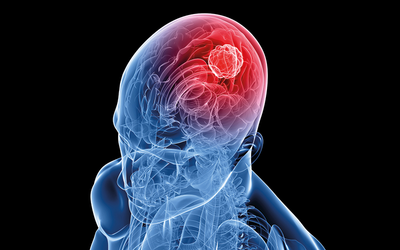
Normally, with a surgical biopsy for a glioma, the surgeon collects a tissue sample, the lab then runs tests to diagnose and, depending on the treatment plan, a second surgical operation may be needed.
However, the researchers from Osaka University have used an advanced DNA-based fluorescence technique that could help bring real-time cancer diagnostics into practice.
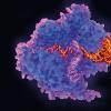
Photo-induced electron transfer is the basis of many DNA-based biosensors. Understanding of the kinetics of this process is based on the average behaviour of many molecules, known as ensemble measurements.
Shuya Fan, lead author, said: “Such measurements obscure the single-molecule behaviour that’s foundational to the kinetics of electron transfer, but our research clears this obscurity. We used fluorescence correlation spectroscopy to measure transient fluorescence patterns and in so doing uncovered single-molecule chemistry that will advance diagnostic applications.”
The scientists used the corresponding fluorescence blinking to detect an mRNA glioma point mutation in cultured cells.
The researchers’ approach is compatible with real-time glioma diagnosis during a surgical biopsy.
Image Credit | iStock