Non-alcoholic fatty liver disease is becoming the most common chronic liver disorder in developed countries.
Histological analysis of liver tissue is the only widely accepted test for diagnosing and distinguishing different stages of the disease.
However, this technique provides only two-dimensional images of the liver tissue in low resolution and overlooks potentially important 3D structural changes.
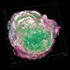
Scientists have now generated 3D geometrical and functional models of human liver tissue for different disease stages. They reveal new critical tissue alterations, providing new insights into pathophysiology and contributing to high-definition medical diagnosis.
Conventional histological analysis of liver tissue is the gold standard for diagnosing disease progression, but it has several potential disadvantages: the low-resolution 2D images of liver tissue may only allow a semi-quantitative evaluation and it can be subjective, since it depends on interpretation.
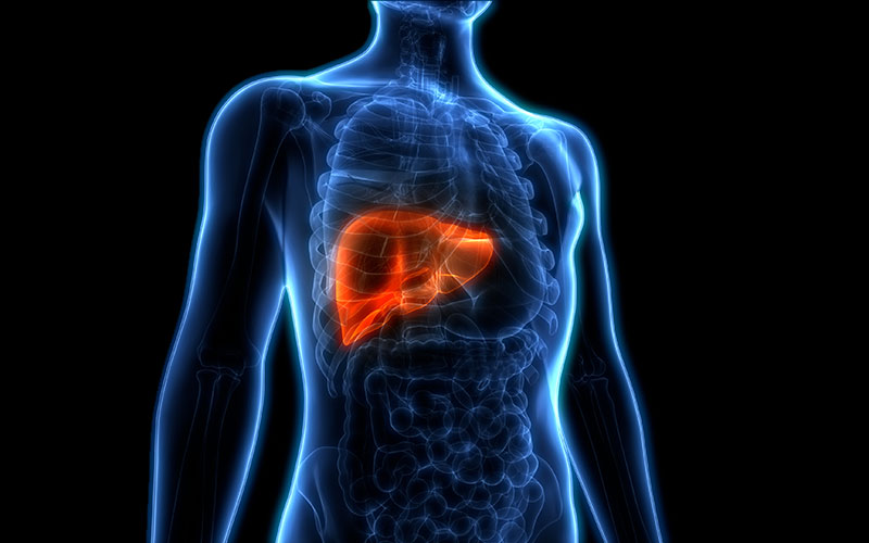
Image credit | Shutterstock