News

AddToAny

Google+ Facebook Twitter Twitter

3D images of tumours in subcellular resolution

Scientists at the Francis Crick Institute have developed an imaging technique that allows simultaneous 3D visualisation of multiple tissue types, providing one of the most comprehensive views of conditions, such as cancer, to date.

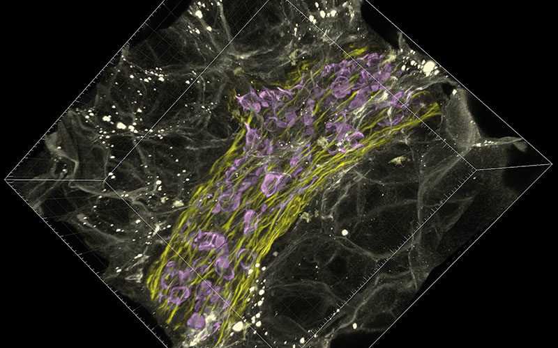
Details of their new method called “fast light microscopic analysis of antibody stained whole organs”, or FLASH, highlight how the technique can be used to examine intact tissue and even whole organs without harm to the sample integrity.

Most imaging methods require ultra-thin samples of just a few micrometres thick, which may not be reflective of the rest of the sample and cannot provide insight into the spatial interactions between different cell types. For example, how cancer cells and immune cells interact.

Over a period of about two years, the team of scientists trialled different combinations of reagents, techniques and timings, until they managed to find a process that allowed them to capture the detailed 3D images, while leaving the sample or organ intact for further analysis.

Hendrik Messal, first author and postdoctoral fellow at the Netherlands Cancer Institute, said: “The concept of 3D imagery is fast evolving and researchers are always testing the boundaries of what we can see using the latest technologies and techniques.

“FLASH is a fast and easy method that can be quickly adopted by researchers familiar with light microscopy techniques and, importantly, it doesn’t damage the tissue, which allows for additional diagnostic tests.”  

go.nature.com/36t66Ct


Related Articles

Medical Science Laboratory with Diverse Multi-Ethnic Team of Microbiology Scientists Have Meeting on Developing Drugs, Medicine, Doing Biotechnology Research-CREDIT_istock-1293772951

Equity, diversity and inclusion for all

Jemma Shead, a Senior Biomedical Scientist at Synnovis and IBMS EDI Working Group member, on how and why you should get involved with EDI.

adeno associated virus capsid-Image Credit | Science Photo Library - c0142837

Examining the medical mystery of child hepatitis outbreak

A study has shed light on an unexpected wave of severe acute hepatitis cases in 2022, amid the backdrop of the ongoing global COVID-19 pandemic.

clostridioides difficile bacteria-Image Credit | Science Photo Library - c0016337

Faecal microbiota transplant for C. diff

In the first comprehensive US evidence-based guideline on the use of faecal microbiota-based therapies for gastrointestinal disease, the American Gastroenterological Association recommends faecal microbiota transplant (FMT) for most patients with recurrent Clostridioides difficile (C. diff) infection.

pseudomonas aeruginosa bacteria Image-Credit | Science Photo Library - f0381226

Tackling priority pathogen with phages

A new study describes the use of phage therapy to eradicate multidrug-resistant Pseudomonas aeruginosa infections in vivo with important new implications for antibiotic resistance.

Top