They may only be small collections of cells at present, but neurological surgeon Ben Waldau believes that brain organoids could have a big impact in the future.

A haughty scientist growing human brains in a laboratory full of bubbling beakers is a classic horror film trope, so the fact that brains are actually being grown in labs around the world might trigger some alarm. The reality is more prosaic, though, as the would-be brains – more usually called cortical spheroids, neural organoids or whole brain organoids – are mostly just small collections of cells, not more than a few millimetres across, that sit in petri dishes. Even so, these organoids are throwing open potential avenues of research, not to mention therapeutic possibilities, that stretch far beyond the realms of creaky old science fiction.
All about Ben
-
Trained at Ruprecht-Karls University, Heidelberg, Germany, 2002
-
Internship in general surgery, then a residency in neurosurgery, at Duke University Medical Center, Durham, North Carolina, 2004-10
-
Fellowship in endovascular neurosurgery, University of Florida, 2010-11
-
Fellowship in neural stem cell research, at the Center for Regenerative Therapies, Dresden, Germany, 2011-12
-
Now Associate Professor in neurological surgery at University of California Davis Medical Centre
- Specialises in open cerebrovascular and endovascular treatment of cerebral aneurysms and arteriovenous malformations.
Inspiration
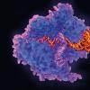
A prime example of this burgeoning area of research emerged in March from the University of California Davis Medical Centre in Sacramento, where a team headed by neurological surgeon Ben Waldau revealed it has been nursing an organoid that has developed its own blood vessels.
Ben’s inspiration for the research came from observing colleagues at Davis.
“I collaborate with the liver disease team,” he says, “and they had developed a protocol for turning induced pluripotent stem cells (IPSCs) into endothelial cells that line the inside of blood vessels. Since we work next to each other I was getting interested in their research and wondered if the same thing that works with the liver would also work with the brain.”
The donor cells came from a patient having a routine surgical procedure. During surgery he opened the dura, the thick layer around the brain, and took a sample of stem cells. One group of these IPSCs was set aside to become the whole-brain organoids, and another to be differentiated into endothelial cells. “We used the protocol to turn the patient’s IPSCs into endothelial cells,” says Ben, “and we let them grow in separate dishes until they were matured. Then we combined them, coating the organoids with the endothelial cells.”
Nature then took its course. “Human brains have their own programme of development,” explains Ben. “Blood vessels come from the outside, from a vascular plexus that’s around the outer brain, and they keep on growing into the brain during development. By coating the organoids with endothelial cells, we tried to mirror this normal human development. The endothelial cells have very good self-organising capacities. Initially they just sit on the organoids, but the next day you look into the microscope and they are forming tubular structures. It is very interesting how they organise themselves.”
The results
The in vitro results were striking, to say the least, and were subsequently tested in vivo when a tiny vascularised organoid was transplanted into the brain of mouse. The organoid not only survived but also developed further blood vessels that grew deeper into the brain of the mouse.
“The in vitro model had shown that the blood vessels were growing into the organoid, so it wasn’t vital to put the organoid into the mouse, but with the rodent model we wanted to see if the blood vessels were also able to survive in vivo,” says Ben. “We wanted to see if it would work because we potentially want to look at future at transplantation experiments where, for example, we can help stroke survivors.”
These organoids are still only a few millimetres across, but a key implication of their vascularisation is that it could be the means for them to grow larger. Ben, however, is not getting too excited at this prospect. “We are just showing proof of concept, that we could vascularise organoids. Whether they can grow bigger, we don’t know yet. If they did, it would certainly aid our research and they might survive better in transplantation.”
Transplantation
If the ultimate vision is to transplant lab-grown brain matter into patients, Ben is not about to get ahead of himself with the idea. “I think that is many years off,” he says. “There is a big regulatory process and we have to be very careful as there are many potential adverse events. First, this has to be approved by the FDA before anything else, and for that we need to collect way more data to prove it is safe. We also have the address concerns, such as the purity of the cells. I think it is up to 10 years down the line before the human transplantation experiments.”
In theory, though, the increasing sophistication of whole brain organoids and their newly developed blood vessels could be a gateway to treating all sorts of neurological disorders, though Ben remains determinedly realistic. “Some conditions, like Alzheimer’s, are so diffuse I am not sure what matter we might be able to rescue there. Other conditions, such as Parkinson’s, are located deep in the mid brain, so single cell infusions with a needle may still be better. What comes to my mind, because I am a vascular surgeon, is stroke. This is limited to certain parts of the brain, often in the cortex at the surface of the brain, which is easier to reach. I think the initial focus will be on replacing lost tissue here.”
Infancy
Instead, Ben’s more immediate focus is on the potential of his work for further study of the brain. “An intrinsic benefit to a vascularised organoid in a dish may be that you can study the blood-brain barrier. Currently this is studied a lot in animals, but here we have a potential human model. For example, we could see whether pharmacological agents are able to cross the blood-brain barrier. We could also look at conditions such as epilepsy or ruptured aneurysms for the first time in a more complex system. It is in its infancy, but in a few years we may be able to study the blood-brain barrier in organoids rather than a rodent model. That is what makes me excited.”